Lecture on Ultrasound Guided PRP Injections
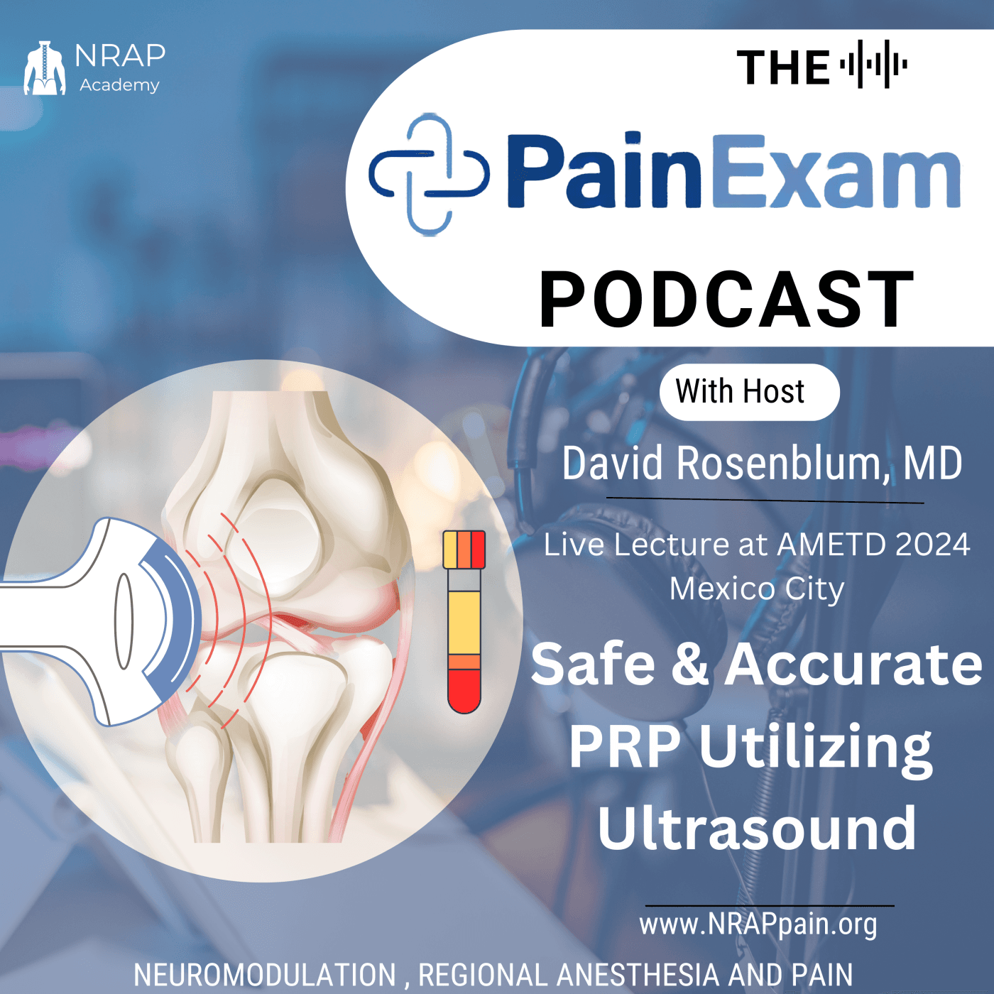
Dr. Rosenblum serves at AMETD's 2024 Conference as faculty and discusses the safe and accurate usage of Ultrasound to Guide PRP injections
Discussed in this lecture:
Knee, Hip, Shoudler, Ligament and Tendon Targets, the ultrasound technique, the evidence for PRP and controversy.
Platelet rich plasma in treatment of patients with idiopathic carpal tunnel syndrome
- Purpose: “To study the effect of PRP injection in treatment of mild to moderate idiopathic carpal tunnel syndrome.”
- Method: RCT in patient who suffered from mild to moderate carpal tunnel syndrome. 98 patients were randomly divided into two groups: group 1 (n=49) had patients who received ultrasound guided PRP injection and group 2 (n=49) had ultrasound guided corticosteroid injection. Outcomes were assessed using the visual analog scale, the Boston carpal tunnel syndrome questionnaire, electrophysiological findings in sensory and motor studies of the median nerve, and morphological changes of the median nerve that were observed via ultrasound. Outcomes were assessed at baseline before the injections, after 1 month of receiving the injection, and after 3 months of receiving injection.
- Results/ Conclusion: In both groups, when compared to baseline, there was improvement in clinical manifestations, the electro diagnostic examination parameters of the median nerve, and the median nerve cross-sectional area at one and three months post injection. PRP injection was also found to be better than the local steroid injection in the improvement of clinical manifestations as well as the motor nerve conduction velocity, the sensory latency, and the motor nerve sensory conduction - these were seen at the 3 month mark.
- https://link.springer.com/article/10.1007%2Fs10067-019-04719-7

Treatment
- Non-surgical treatments include wearing a wrist splint, nonsteroidal anti-inflammatory drugs, and cortisone injections.
- Despite the availability of conservative therapies, their efficacy is usually short-lived.
- Surgery is recommended when carpal tunnel syndrome does not respond to conservative measures.
- The goal of surgery is to increase the size of the tunnel in order to decrease the pressure on the nerves and tendons that pass through the space.
Ultrasound Guided PRP Injections of the Upper Extremity - Shoulder
Biologics on Rotator Cuff Tears
- PRP not only inhibits the inflammatory effects of interleukin 1β (IL-1β) but also enhances TGF-β production
- Increased concentration of IL-1β is significantly correlated with rotator cuff tendon degeneration;
- TGF-β enhances rotator cuff tendon repair strength
- Controlled clinical studies using different autologous PRP formulations have provided controversial results.

Ultrasound Guided PRP Injections for Partial Rotator Cuff Tears
20 patients with symptomatic partial rotator cuff tears were treated with ultrasound guided platelet rich plasma injection.
Before and after the injection of platelet rich plasma scoring was done with visual analogue score
Constant shoulder score, and UCLA shoulder score at 8 weeks and third month.
A review ultrasound was performed 8 weeks after platelet rich plasma injection to assess the rotator cuff status
Results:
Our study showed statistically significant improvements in 17 patients in VAS pain score, constant shoulder score and UCLA shoulder score.
No significant changes in ROM were noted when matched to the contra-lateral side (P < 0.001) at the 3 month follow-up.
The study also showed good healing on radiological evaluation with ultrasonogram 8 weeks after platelet rich plasma injection
Sodium hyaluronic and PRP for partial thickness rotator cuff tears.
- Purpose: The aim of this study was to evaluate the combined used of sodium hyaluronate and PRP in the treatment of small to medium partial thickness rotator cuff tears
- Method: N=200 Double blinded randomized trial. Partial thickness rotator cuff tears were detected by clinical exam and MRI and they were included in the study. Patients were subsequently randomly assigned to receive subacromial injection of either saline, SH, PRP, or SH + PRP once a week for four weeks. Primary outcome measures was the Constant score and secondary outcome included ASES (American shoulder and elbow surgeons) and VAS score. All of these measures were checked pre-treatment and then at 1, 3, 6, and 12 months post treatment. MRI was also used to evaluate how the rotator cuff changed after 1 year post treatment.
- Results/ Conclusion: “PRP and SH+PRP group had significantly higher Constant score and ASES score after the treatments… MRI results showed that the tear size significantly decreased in both the PRP & the SH + PRP group, especially the SH + PRP group.” The study provided evidence showing the efficacy of PRP injections when it came to healing small to medium partial thickness rotator cuff tears. Also, combination of SH and PRP give better results than either of them alone.
- https://journals.lww.com/acsm-msse/Fulltext/2019/02000/Sodium_Hyaluronate_and_Platelet_Rich_Plasma_for.1.aspx
Glenohumoral Joint Injection Glenohumoral Joint Biceps TendonBiceps Tendon Subscapularis Tendon Supraspinatous Tendon
Subacromial Bursa
- 25G 1.5 inch needle or 25 G 3 1/2 inch spinal needle
- 3 to 4ml injectate
Subacromial Bursa
- 25G 1.5 inch needle or 25 G 3 1/2 inch spinal needle
- 3 to 4ml injectate
DeQuervaine’s Tendosynovitis
Thickening of and accumulation of mucopolysaccharide in the sheath of
- Abductor Pollicis Longus Tendon
- Extensor Pollicis Brevis Tendon
The Extensor Reticnaculum is a fibrous band attached to the radius and prevents bowstringing of the extensor tendons off the dorsum of wrist
Ultrasound Guided PRP Injections for De Quervaine’s Tendosynovitis
Symptoms
Pain near the base of the thumb
Swelling near the base of the thumb
Difficulty moving the thumb and wrist when grasping or pinching during activities
A "sticking" or "stop-and-go" sensation in your thumb when moving it
Signs Positive Finklestein’s Test: the examiner grasps the thumb and ulnar deviates the hand sharply. if sharp pain occurs along the distal radius (top of forearm, close to wrist; see image), de Quervain's tenosynovitis is likely.
Eichhoff's test: the Eichhoff's test is typically described as the examiner grasping and ulnar deviating the hand when the person has their thumb held within their fist. If sharp pain occurs along the distal radius, De Quervain's tenosynovitis is suspected but more likely to produce false positive
DeQuervaine’s Tenosynovitis
Stenosis tenosynovitis
- Scan the radial aspect of the wrist.
- Extensor pollicis brevis and abductor pollis longus tendons are both involved because they share a common tendon sheath.
- Caused by overuse movements that require repetitive extension and abduction of the thumb.
- Superficial Injection
- Skin atrophy and hypopigmentation may occur with steroids
DeQuervaine’s
Treatment
Aim of Study: To evaluate the effects of PRP in the treatment of DQVD in comparison with corticosteroid (CS) injection.
Subjects and Methods: 40 hands of 35 patients were included. 20 hands received single CS injection (group I) and the other 20 hands were injected with PRP (group II). Patients were evaluated using Visual analogue scale (VAS), Jebsen Hand Function Test (JHFT), the shortened disabilities of the arm, shoulder and hand questionnaire (Quick DASH), and the examination of the contents of the first dorsal compartment by MSUS at baseline, one and six months after the injection.
Healthy side of patients were considered as control group (30 hands).
Results: Overall, 31 females and 4 males were included with 20 hands in each group. The CS group had better pain relief, hand function tests and ultrasonograghic findings at 1 month, but at 6 months follow-up, the PRP group had statistically significant better pain relief, hand function tests and US findings than CS group.
Ultrasound Guided PRP Injections for Lateral Epicondylitis
Lateral Epicondylitis
- Extensor carpi radialis brevis (ECRB) is the most commonly affected muscle
Treatment
Medial Epicondylitis
Posterior Elbow
- A bright (echogenic) triangular or oval nerve distal to the elbow
- Joins the ulnar side of the ulnar artery in the forearm where it is surrounded by the FDS and FDP and flexor carpi ulnaris (FCU) muscles.
- In the distal forearm the ulnar nerve will lie adjacent to the FCU tendon
Posterior Elbow
- A bright (echogenic) triangular or oval nerve distal to the elbow
- Joins the ulnar side of the ulnar artery in the forearm where it is surrounded by the FDS and FDP and flexor carpi ulnaris (FCU) muscles.
- In the distal forearm the ulnar nerve will lie adjacent to the FCU tendon
Ultrasound Guided PRP Injections for Trigger Finger
- Thickened flexor tendon in distal palm
- Abnormal gliding and locking of Tendon within Sheath
- Tendon caught at edge of first annular pulley
- Catching during active flexion-extension
- Stiff, tender, palpable nodule
- Associated conditions RA, Gout
Trigger Finger
- 25G 1.5 inch needle or smaller
- Low volume
- Feel Tendon Sheath get Distended with injection
- Out of plane approach may be necessary but use caution to avoid tendon rupture
Ultrasound Guided PRP Injections for Achilles Tendinopathy
- Although many of the retrospective studies suggested an advantage of using PRP, the higher level of evidence studies do not support a significant efficacy.
- A large randomized trial by Keene et al found no evidence that PRP injections improved muscle tendon function, patient reported function, pain, goal attainment, or quality of life in patients.
- The hypothesized benefits of PRP plasma in tendon injury healing, based on encouraging findings in laboratory studies did not translate into a detectable patient benefit.
Effects of platelet rich plasma on healing tissues in acute ruptured Achilles tendon: a human immunohistochemistry study
- Purpose: To investigate the response of ruptured Achilles tendons that are treated with PRP.
- Method: Achilles tendon tissue biopsies were taken from 20 patient with ruptured Achilles tendon's via ultrasound guided needle biopsies. These biopsies were taken from the healing area of the Achilles tendon's six weeks after treatment with PRP or after treatment with the placebo (control group) - 10 people in each group. These samples were imbedded in paraffin wax and stained - looked at type 1 and 3 collagen, CD45 lymphocyte, blood vessels and cellular proliferation.
- Results/ Conclusion: Increased cellularity and glycosaminoglycans (crucial role in regulation of cell growth, proliferation, and wound repair) in the PRP treated tendon group than the control group. Modified Bonar score was lower in the PRP group compared to the placebo control group- indication of improved early tendon healing.
- “locally applied PRP enhanced the maturity of the healing tendon tissues by promoting better collagen-1 deposition, decreased cellularity, less vascularity, and higher glycosaminoglycan content when compared with control samples”
- https://www.thelancet.com/journals/lancet/article/PIIS0140-6736(15)60334-8/fulltext
Evidence for the use of PRP in chronic mid-substance Achilles tendinopathy: A systematic review with meta-analysis
The use of PRP injections in the management of chronic mid-substance Achilles tendinopathy is increasing, but the efficacy is highly debated
Randomized controlled trials with a high level of methodological quality show no clear additional value of PRP. Whereas studies favoring PRP seem to have a higher risk of bias profile.
PRP injections show no clear additional value in the management of chronic midsubstance AT, therefore the widespread use of PRP is questionable.
Platelet rich plasma treatment of non-insertional Achilles tendinopathy in a two case series: no significant difference in affect between leukocyte rich and leukocyte poor PRP
- Purpose: Comparison of two case series to assess 1) the effect of PRP on chronic Achilles tendinopathies (CAT) and 2) if there is any difference in effect between leukocytes rich PRP and leukocyte poor PRP in the treatment of CAT.
- Method:84 patients who failed to to show progress with “normal” treatment of chronic Achilles tendinopathy for at least 6 months were treated with Leukocyte rich PRP (36 patients) or Leukocyte poor PRP (48 patients). Patients in both groups were asked to stop NSAIDs - 1 week before treatment and for 2 months after treatment. Patients were also kept NWB for 2 wks afterwards, but ankle movement was encouraged. VAS at rest and with activity was used for pain intensity. CAT severity gauged using VISA-A scale. Both of these were self reported at baseline and then at 2 months from initiation of Tx.
- Results w. LR PRP group = VAS at rest declined from an average to 4.0 to 1.1, VAS with activity declined from 7.3 to 3.4 and then further to 1.8 when checked at closer to 37 months. VISA-A increased from 45.4 to 56.5 - 26/ 27 patients who were satisfied stated they would choose PRP again (out of 36 patients total)
- Results w. LP PRP group = VAS at rest declined from an average of 4.2 to 1.1, VAS with activity declined from 7.8 to 4.8 and further to 3.6 at about 37 months. VISA-A increased from 29.7 to 44.7 on average - 11/19 patients who were satisfied would choose PRP again.
- ”PRP seems to be a possible treatment when all other treatment regimens have failed, with a reasonably high probability of reaching MCIC (minimal clinically important change). The choice of either LR-PRP or LP-PRP seems to be up to personal preference as there were no significant differences between patients treated with LR-PRP and LP-PRP.”
- •https://www.ncbi.nlm.nih.gov/pmc/articles/PMC6460817/
Treatment
- Numerous methods have been advocated for treating plantar fasciitis, including rest, nonsteroidal anti-inflammatory medication, night splints, foot orthosis, stretching protocols, and extracorporeal shock wave therapy.
- Corticosteroid injections
- Surgery in severe cases
- Platelet-rich plasma (PRP) has proven to be a safe therapeutic option in the treatment of tendon, muscle, bone, and cartilage injuries.
Ultrasound Guided PRP Injections for Plantar Fasciitis
- According to double-blind randomized trial by Peerbooms et al, an injection of PRP improves the outcome of patients with chronic plantar fasciitis more so than corticosteroid injections.
- The primary outcome parameter was pain and secondary parameters were function and quality of life.
- Patients in the PRP group showed significantly lower pain and disability scores than patients in the control group after adjusting for baseline differences.
- According to Gonnade et al10 in their recent article, previous observational studies and a few randomized clinical trials on plantar fasciitis have concluded that PRP is an effective therapy in chronic cases, but still there is controversy owing to a lack of level 1 evidence
- The most recent study by Mahindra et al18 found that PRP and cortisone are better than placebo, but at 3 months of follow-up, PRP injection was significantly better than corticosteroid injection.
- Peerbooms, J. C., Lodder, P., den Oudsten, B. L., Doorgeest, K., Schuller, H. M., & Gosens, T. (2019). Positive Effect of Platelet-Rich Plasma on Pain in Plantar Fasciitis: A Double-Blind Multicenter Randomized Controlled Trial. The American Journal of Sports Medicine, 47(13), 3238–3246. https://doi.org/10.1177/0363546519877181
Platelet rich plasma has better long-term results than corticosteroids or placebo for chronic plantar fasciitis: a randomized controlled trial
- Purpose: “To compare the pain and functional outcomes of PRP with CS and placebo injections for the treatment of chronic plantar fasciitis.”
- Method: 3 arm randomized controlled trial - 90 patient’s: 30 PRP, 30 steroids, 30 placebo. Followed until 18 months post injection. VAS, Roles Maudley score, and the mean Short Form 12 score were used for tracking outcomes.
- Results/ Conclusion: VAS showed improvement in all groups from baseline to 18 month follow up with steroids showing significant improvement to PRP in the short term but long term the results for PRP were better than steroids. The PRP group showed better results long term compared the steroid group when looking at pain, function, and general health. “Both PRP and CS are safe and effective treatment options for chronic plantar fasciitis, showing superior results to placebo treatment-the longer term results and less reinfection and/or surgery rate of PRP makes it more favorable as an injection option than steroid injections.
- Level 2 evidence
- https://linkinghub.elsevier.com/retrieve/pii/S1067-2516(18)30282-5
Platelet rich plasma versus corticosteroids injections for plantar fasciitis: a comparative study
- Purpose: PRP vs . Cortisone for chronic planter fasciitis resistant to traditional non-operative management
- Method: 60 heels included in study, randomized into PRP and steroid injection group. Outcomes were measured using Roles Maudsley score, VAS, and American Ortho. Foot and Ankle Society score - checked at pre-treatment, & then 3,6, & 12 months post injection
- Results/ Conclusions: at 3 and 6 months, PRP was found to be as effective as a steroid injection for treating plantar fasciitis - at 12 months the PRP group showed statistically significant improvement compared to the steroid injection group (RM, VAS, and AOFAS scores were all better)
- https://www.sciencedirect.com/science/article/abs/pii/S0958259215000838?via%3Dihub
Ultrasound Guided PRP Injections for Osteoarthritis of the Hip
Hip Injection
- A total of 4 RCTs were identified comparing PRP with HA, one of which also compared it against an injection of both HA and PRP.
- In all of these studies, PRP was not found to be superior to HA. There was no significant difference between the two for the treatment of hip OA.
- •https://doi.org/10.1007/s11845-020-02388-z
The use of ultrasound guided PRP injections in the treatment of hip osteoarthritis: a systematic review of the literature (level one evidence, a systematic review of level one studies -2018)
- Purpose: Aim of this study is to determine whether ultrasound guided PRP injection has a role in improving clinical outcomes in patients with hip OA.
- Method: Looked at 3-level one evidence studies (RCTs) that met the inclusion criteria - total of 254 patients in the 3 studies, 115 received PRP (61 males/ 54 females), with 31 patients who got the PRP and control. All 3 studies had HA as the control and all injections were given under US. They had varying degrees of Hip OA based on the Kellgren and Lawrence (KL) classification.
- Results/ Conclusion: “Outcome scores show an improvement of symptoms and function maintained up to 12 months following PRP injections.” This article suggests that ultrasound guided a PRP injections may be an effective way of delivering long-term and clinically significant pain reduction in people with osteoarthritis. It also may lead to an improvement in function.
- https://pubmed.ncbi.nlm.nih.gov/30763018/
Ultrasound guided injection of PRP and HA, separately and in combination for hip osteoarthritis (a randomized controlled study)
- Purpose: Compare the therapeutic efficacy of autologous PRP versus hyaluronic acid versus PRP +HA in the treatment of hip OA.
- Method: 111 Patients (ages 18-65), with history of having outpatient hip surgery, and hip OA at time of study. They were put into 3 groups & each received 3 injections, each one week apart - 44 with PRP, 31 with PRP+HA and 36 with HA. All patients scored above 20mm on VAS. Outcomes measured via VAS at 2,6, and 12 months after treatment and also using the Harris Hip Score, WOMAC, and concentration GF in PRP + its correlation with clinical outcomes. Clinical outcomes were evaluated by people blinded to the type of treatment the patients received.
- Results/ Conclusion: At all follow ups the PRP group had the lowest VAS score. At 6 months follow up: PRP’s VAS was 21 on average, PRP + HA VAS was 35, and HA VAS score was 44. WOMAC score for PRP group was better at 2 and 6 month follow up, but not at 12 month. Use of PRP + HA did not lead to significant improvement in pain.
- https://journals.sagepub.com/doi/10.1177/0363546515620383
Greater Trochanter Pain Syndrome Treatment
- If conservative measures fail then progressively more invasive treatment options including shockwave therapy, corticosteroid injections (CSI), PRP and surgery may be required
- Surgical treatment for GTPS is reserved for recalcitrant cases Procedures are dependent on underlying pathology but may involve lengthening or release of the ITB and fascia lata, gluteal tendon tear repair, minimally invasive endoscopic bursectomy, or open reduction trochanteric osteotomy.
Knee PRP Targets Injection
- Lateral Collateral Ligament
- Lateral Meniscus
- Iliotibial Band
- Housemade’s Knee
- Housemade’s Knee
- Housemade’s Knee
- PainExam App is ready for iphone

- Pain Management Board Prep migrated to NRAPpain.org
- AnesthesiaExam Board Prep migrated to NRAPpain.org
- PMRExam Board Prep migrated to NRAPpain.org


References
https://rapm.bmj.com/content/rapm/early/2024/07/16/rapm-2024-105593.full.pdf
Disclaimer
Disclaimer: This Podcast, website and any content from NRAP Academy (NRAPpain.org) otherwise known as Qbazaar.com, LLC is for general informational purposes only and does not constitute the practice of medicine, nursing or other professional health care services, including the giving of medical advice, and no doctor/patient relationship is formed. The use of information on this podcast or materials linked from this podcast is at the user’s own risk. Professionals should conduct their own fact finding, research, and due diligence to come to their own conclusions for treating patients. The content of this podcast is not intended to be a substitute for professional medical advice, diagnosis, or treatment. Users should not disregard or delay in obtaining medical advice for any medical condition they may have and should seek the assistance of their health care professionals for any such conditions.