Table of Contents
- Lumbar Facet Joint Anatomy and Clinical Presentation
- Facet-Mediated vs. Discogenic vs. Radicular Pain
- Facet Joint Pain Treatment: Medial Branch Blocks and RFA
- Peripheral Nerve Stimulation for Facet-Mediated Pain
- Platelet-Rich Plasma (PRP) for the Facet Joint
- How Common Is Facet-Mediated Low Back Pain?
- Post-Procedure Care and Expected Recovery
- Video: Ultrasound Technique Demonstration
- Keep Learning
- NRAP Academy Courses
- Looking for a Pain Specialist?
- FAQs
Low back pain is one of the most common complaints in any pain management practice, and lumbar facet joints are a leading — and frequently under-diagnosed — pain generator behind it. For physicians preparing for pain medicine boards, distinguishing facet-mediated pain from discogenic and radicular pain, and knowing the full ladder of facet joint pain treatment options, is classic exam territory and classic clinical territory alike. This guide walks through the clinically relevant anatomy, the diagnostic algorithm, and the evidence behind radiofrequency ablation, peripheral nerve stimulation, and platelet-rich plasma (PRP) as facet joint pain treatment options.
Note: caudal epidural steroid injections are not covered here. That technique targets the epidural space for lumbosacral radicular pain and spinal stenosis, a different pain generator from the facet joint — see our separate post on caudal epidural steroid injections for lumbar radiculopathy and spinal stenosis for that technique and the evidence behind it.
In this post:
- Lumbar Facet Joint Anatomy and Clinical Presentation
- Facet-Mediated vs. Discogenic vs. Radicular Pain
- Facet Joint Pain Treatment: Medial Branch Blocks and RFA
- Peripheral Nerve Stimulation for Facet-Mediated Pain
- Platelet-Rich Plasma (PRP) for the Facet Joint
- How Common Is Facet-Mediated Low Back Pain?
- Post-Procedure Care and Expected Recovery
- Video: Ultrasound Technique Demonstration
- Keep Learning
- NRAP Academy Courses
- Looking for a Pain Specialist?
Lumbar Facet Joint Anatomy and Clinical Presentation
The lumbar facet joints (zygapophyseal joints) are paired synovial joints formed by the inferior articular process of the vertebra above and the superior articular process of the vertebra below. Each joint is a true diarthrodial joint with a synovial lining, hyaline cartilage, and a fibrous capsule — which is exactly why facet joints can become arthritic and painful in the same way any other synovial joint can. The capsule and surrounding structures are richly innervated by the medial branches of the dorsal rami, which is the anatomic basis for diagnostic blocks, radiofrequency ablation, and nerve stimulation alike. Understanding this anatomy is foundational to selecting the right facet joint pain treatment for a given patient.
A key board fact: each lumbar facet joint receives dual innervation. The L4-5 facet joint, for example, is innervated by the medial branch of the L3 dorsal ramus (which crosses over the L4 transverse process) and the medial branch of the L4 dorsal ramus (which crosses the L5 transverse process). This is why a diagnostic medial branch block for a given joint always targets two levels, not one — a frequently tested point.
Facet-mediated low back pain classically presents as:
- Axial low back pain, often with referral into the buttock, posterior thigh, and occasionally the groin — but rarely below the knee.
- Pain that worsens with lumbar extension and rotation (e.g., standing up from a flexed position, reaching overhead) and improves with flexion.
- Paraspinal tenderness to palpation over the facet joints, without a focal neurologic deficit.
- Morning stiffness that improves with movement, similar in character to other synovial joint arthropathies.
No single historical or physical exam finding is sensitive or specific enough to diagnose facet joint pain on its own — this is precisely why controlled diagnostic blocks remain the reference standard, a point worth committing to memory for boards.
Facet-Mediated vs. Discogenic vs. Radicular Pain
Distinguishing these three common causes of low back pain is one of the most frequently tested concepts in pain medicine board exams, since the correct facet joint pain treatment pathway depends on getting this distinction right. The table below summarizes the classic distinguishing features.
| Feature | Facet-Mediated Pain | Discogenic Pain | Radicular Pain |
|---|---|---|---|
| Pain distribution | Axial, referral to buttock/posterior thigh, above the knee | Axial, may be diffuse or band-like, occasionally referred to buttock/thigh | Dermatomal, often extends below the knee |
| Aggravating position | Extension, rotation, standing | Flexion, prolonged sitting, axial loading (Valsalva) | Positions that increase nerve root tension (straight leg raise, flexion in disc herniation) |
| Neurologic exam | Normal | Usually normal | May show dermatomal sensory loss, myotomal weakness, or reduced reflex |
| Straight leg raise | Negative | Negative (may be positive if large central herniation) | Often positive |
| Reference-standard diagnostic test | Controlled comparative medial branch blocks | Provocative discography (controversial, rarely first-line) | Selective nerve root block, MRI correlation with exam |
| First-line image-guided facet joint pain treatment | Medial branch block → radiofrequency ablation | Epidural steroid injection (variable benefit), intradiscal therapies | Transforaminal or caudal epidural steroid injection (see our separate post on caudal ESI for radiculopathy and stenosis) |
Facet Joint Pain Treatment: Medial Branch Blocks and RFA
Because no imaging study reliably confirms facet joint pain, the diagnostic pathway for this facet joint pain treatment relies on comparative local anesthetic blocks of the medial branches supplying the suspected joint(s). This is heavily tested material, so it's worth knowing the details precisely.
Technique
Under ultrasound or fluoroscopic guidance, a small volume of local anesthetic (typically 0.3-0.5 mL) is deposited at the junction of the superior articular process and the transverse process — the location where the medial branch courses before it enters the multifidus and interspinous ligament. For the L5 dorsal ramus, the target is the junction of the sacral ala and the superior articular process of S1, since L5 has no true transverse process in the same sense as the other lumbar levels.
Ultrasound guidance for medial branch blocks has become increasingly popular because it avoids radiation exposure, allows real-time visualization of needle advancement relative to the transverse process, and is well suited to an office-based practice, making it a practical facet joint pain treatment for everyday clinic use. The classic ultrasound approach uses a curvilinear probe in a parasagittal orientation to identify the "sawtooth" pattern of the transverse processes, then walks the probe medially to visualize the articular pillar and facet joint complex.
Confirming a true positive response
A single block is not considered diagnostic because of a high false-positive rate (placebo response, spread of local anesthetic to adjacent structures). Board-relevant criteria for a meaningful response include:
- At least 80% pain relief on the treated movements/activities that reproduced the patient's pain, documented immediately after the block.
- Confirmation with a second, comparative block (either a repeat block with the same anesthetic on a separate day, or a differential block using agents with different durations of action, such as lidocaine vs. bupivacaine) to reduce false positives before proceeding to radiofrequency ablation.
- Some protocols accept a single block with strict ≥80% relief criteria given cost and practical constraints, but comparative blocks remain the more rigorous (and more frequently tested) standard.
When radiofrequency ablation (RFA) is indicated
Radiofrequency ablation of the medial branches, the longest-established facet joint pain treatment after a positive diagnostic workup, is indicated only after a patient has met the diagnostic criteria above — it should never be performed as a first-line or "diagnostic" procedure itself. RFA uses heat (typically 80-90°C) generated by a radiofrequency current to create a lesion that denervates the target medial branch, interrupting nociceptive signaling from the joint. Because the medial branch nerve runs along the bone in a roughly cephalad-to-caudad direction, the active tip of the RFA cannula is positioned parallel to the nerve (not perpendicular, as with many other peripheral nerve ablations) to maximize the length of nerve captured within the lesion — another frequently tested procedural detail.
Expected clinical benefit from RFA typically lasts 6-12 months, as the nerve can regenerate over time; repeat ablation is both expected and effective when the original diagnostic workup was sound. Reported complications are generally minor and include transient localized pain, neuritis, and rarely motor weakness if current spreads to a ventral ramus or mixed nerve. For patients who respond well to RFA but want to avoid repeat ablations, or whose pain recurs faster than the typical denervation interval, the two options below are increasingly used as a next-line facet joint pain treatment.
Peripheral Nerve Stimulation for Facet-Mediated Pain
Neurostimulation has emerged as a facet joint pain treatment option for patients with refractory or recurrent symptoms, and it's worth distinguishing two different approaches that are sometimes conflated.
Direct peripheral nerve stimulation (PNS) of the medial branch nerve places a percutaneous lead adjacent to the same medial branch targeted by diagnostic blocks and RFA, delivering low-level electrical stimulation rather than a thermal lesion. The appeal is a reversible, nerve-sparing alternative for patients who have had a clear, reproducible response to medial branch blocks and/or RFA but want to avoid repeated ablations, or whose anatomy or comorbidities make repeat RFA less attractive. The evidence base here is still early: a 2024 case report from the University of Alabama at Birmingham described a patient with chronic facetogenic low back pain from lumbar spondylosis, refractory to facet injections and RFA, who had an L4 medial branch PNS lead placed and reported roughly 80% pain improvement at 60 days.1 The authors were explicit that the supporting evidence for this approach is currently level II–III, and that larger controlled trials are needed before it becomes standard care — a fair characterization of where this facet joint pain treatment option currently stands.
Restorative neurostimulation of the multifidus (marketed as ReActiv8) is a related but mechanistically distinct technology. Rather than targeting the sensory medial branch fibers that carry facet joint pain signals, this system stimulates the motor branch supplying the lumbar multifidus muscle, aiming to restore the neuromuscular control and segmental stabilization that can be lost in chronic mechanical low back pain — a different indication from facet-joint-confirmed pain, though the two populations overlap, since multifidus dysfunction and facet arthropathy often coexist. The pivotal ReActiv8-B trial, a 204-patient, double-blind, sham-controlled study published in Pain in 2021, randomized patients with refractory mechanical chronic low back pain to active versus sham multifidus stimulation.2 The primary responder-rate endpoint at 120 days (57.1% active vs. 46.6% sham) did not reach statistical significance, but secondary outcomes — pain scores, disability, and quality of life — favored active stimulation, and benefit continued to grow over the following year once sham patients crossed over to active stimulation (74% achieving ≥30% pain relief at 1 year). Device-related serious adverse events occurred in about 4% of patients, mostly pocket infections. This is a more rigorously studied technology than direct medial branch PNS, but it is FDA-approved specifically for refractory mechanical low back pain associated with multifidus dysfunction, not as a facet-diagnosis-confirmed therapy — an important distinction when counseling patients and when answering board questions that test the difference between these two neurostimulation approaches.
Platelet-Rich Plasma (PRP) for the Facet Joint
PRP — a concentrate of a patient's own platelets and growth factors prepared from a blood draw — is an emerging, biologically-oriented facet joint pain treatment option, used as an alternative or adjunct to local anesthetic/steroid injection and RFA, particularly for patients who prefer to avoid repeated steroid exposure or ablation.
A 2025 prospective study in Orthopedic Reviews compared CT-guided intra-articular facet joint injections of leukocyte-poor PRP against bupivacaine alone in 78 patients with chronic lumbar facet joint syndrome.3 Both groups had meaningful early pain relief at two weeks, but the PRP group's improvement was more durable — pain scores at one year and Oswestry Disability Index scores at three to six months were significantly better with PRP than with local anesthetic alone. The authors were upfront about the study's limitations: patients selected their own treatment arm rather than being randomized, and the comparison group was relatively small, so they called for larger, multicenter, randomized trials to confirm the finding. That caveat matters clinically — PRP for the facet joint should currently be framed to patients as a reasonable, evidence-informed option with a developing track record, not a proven first-line facet joint pain treatment with the same evidentiary weight as RFA.
The procedural mechanics are the same as PRP used elsewhere in musculoskeletal medicine: a blood draw, centrifugation to concentrate the platelet-rich layer, and injection directly into the facet joint capsule under ultrasound or fluoroscopic/CT guidance to confirm accurate intra-articular placement.
How Common Is Facet-Mediated Low Back Pain?
Lumbar facet joint pain is widely regarded as one of the more common identifiable sources of axial low back pain, particularly in older adults, where degenerative facet arthropathy is prevalent. That said, exactly how often the facet joints are the primary pain generator in a given patient is harder to pin down than it might seem, and published estimates vary considerably across studies. Much of that variability comes down to which diagnostic criteria are used — studies relying on a single, uncontrolled medial branch block tend to report a higher prevalence of facet-mediated pain than studies requiring a comparative or double-block confirmation, since a single block carries a meaningful false-positive rate. Looser diagnostic criteria tend to inflate prevalence estimates, a point worth keeping in mind — and since facet joint pain treatment decisions hinge on an accurate diagnosis, this distinction matters clinically, not just academically.
Facet-mediated pain also becomes more common with advancing age, mirroring the accumulation of degenerative changes within the joint that parallels osteoarthritis elsewhere in the body. It is also important to recognize that facet pain rarely exists in isolation: it can, and often does, coexist with discogenic and radicular pain generators in the same patient, since degenerative changes in the facets, discs, and neural foramina tend to progress together. This overlap is part of why a structured diagnostic approach — history, exam, and, when appropriate, controlled blocks — matters more than assuming a single pain generator from imaging alone. Degenerative facet changes are extremely common on imaging even in people with no back pain at all, so imaging findings by themselves do not establish the facet joints as the cause of a given patient's pain.
Post-Procedure Care and Expected Recovery
After a medial branch block or radiofrequency ablation, patients are typically observed briefly and then discharged home the same day, since both are outpatient procedures performed with local anesthesia (and often mild sedation) rather than general anesthesia. Mild soreness at the injection site for a day or two afterward is common and expected.
For diagnostic medial branch blocks specifically, patients are generally asked to track their pain closely in the hours immediately following the procedure, often using a pain diary or a structured activity-based assessment that asks them to attempt movements that previously reproduced their pain. This matters because the diagnostic value of the block depends on documenting the degree and duration of relief while the local anesthetic is still active.
After radiofrequency ablation, it is fairly common for patients to experience a temporary increase in pain, or a new neuritis-type discomfort, for one to a few weeks as the ablated nerve degenerates. This should be anticipated and discussed with patients beforehand so it isn't mistaken for treatment failure; the expected benefit of RFA typically becomes apparent only once this window has passed.
PNS lead placement (whether at the medial branch or the multifidus motor branch) is typically done as a staged process — a temporary trial period to confirm meaningful benefit before a permanent system is implanted — and patients are counseled on activity restrictions around the incision site during the healing period. For PRP as a facet joint pain treatment, soreness at the injection site for a few days is expected and is part of the intended local inflammatory response, and meaningful benefit, consistent with PRP's mechanism elsewhere in the body, tends to build gradually over several weeks rather than appearing immediately.
Video: Ultrasound Technique Demonstration
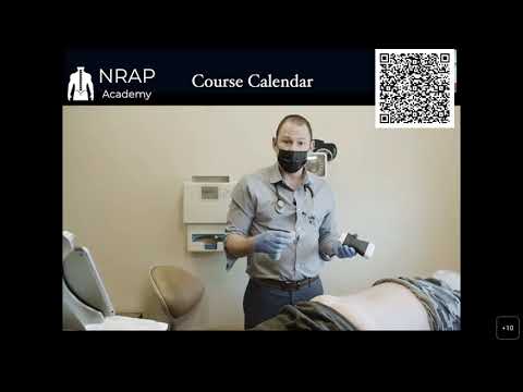
The video below is a multi-procedure ultrasound-guided spine demonstration from NRAP Academy that includes a segment on lumbar facet joint/medial branch targeting — one facet joint pain treatment approach covered above — alongside several other ultrasound-guided spine and nerve procedures. Watch for the probe positioning over the articular pillars for facet targeting.
Keep Learning
For a deeper dive into the evidence behind lumbar facet joint interventions and facet joint pain treatment guidelines, listen to the PainExam Podcast episode "Consensus Statement on Facet Joints Reviewed!", where Dr. Rosenblum discusses the multispecialty consensus practice guidelines on facet joint interventions. For the sources cited above on nerve stimulation and PRP, see the full studies: Strawn et al. on medial branch PNS,1 Gilligan et al. on the ReActiv8-B restorative neurostimulation trial,2 and Baltzer et al. on PRP for lumbar facet joint syndrome.3 You can also find more ultrasound demonstrations on the NRAP Academy YouTube channel. For related high-yield topics, see our posts on cervical radiculopathy and C6 nerve root injections, occipital neuralgia and lesser occipital nerve block, and our companion post on caudal epidural steroid injections for lumbar radiculopathy and spinal stenosis.
1. Strawn LD Jr, Glasser K, Sellers A. Peripheral Nerve Stimulation of Lumbar Medial Branch Nerves for the Treatment of Facetogenic Low Back Pain. Southern Pain Society, 2024.
2. Gilligan C, et al. (ReActiv8-B investigators). An implantable restorative-neurostimulator for refractory mechanical chronic low back pain: a randomized sham-controlled clinical trial. Pain. 2021;162(10):2486-2498. doi:10.1097/j.pain.0000000000002258
3. Baltzer AW, Enneper J, Baltzer LM, Godde G. Platelet Rich Plasma for the Therapy of the Lumbar Facet Joint Syndrome: A Prospective Study About CT-Guided Facet Joint Injections With PRP Compared to Local Anesthetics. Orthopedic Reviews. 2025;17. doi:10.52965/001c.141416
NRAP Academy Courses
If you're preparing for pain medicine boards or want structured, hands-on ultrasound training in spine and musculoskeletal interventions, see the PainExam Pain Management Board Review and Refresher Course and the Ultrasound Training programs at NRAP Academy.
Looking for a Pain Specialist?
Looking for a pain specialist? Visit RosenblumPain.com or call (718) 436-7246. Patients outside the NYC/Brooklyn area can also book a virtual second-opinion consultation through RosenblumPain.com.
Disclaimer: This content is provided for general educational and informational purposes only and is intended for healthcare professionals, trainees, and patients seeking general information. It is not medical advice, does not constitute a diagnosis or treatment recommendation for any individual, and does not create a physician-patient relationship. Treatment decisions must always be individualized by a qualified, licensed healthcare provider based on a patient's specific history, examination, and current evidence-based guidelines. Dr. David Rosenblum, NRAP Academy, and PainExam make no warranties as to the completeness or applicability of this information and assume no liability for actions taken based on it. If you are a patient, please consult your own physician before making any medical decisions, and seek immediate medical attention for any urgent or emergency symptoms.