Table of Contents
- Cervical Radiculopathy: Pathophysiology and Classic Patterns
- C5–C8 Dermatome, Myotome, and Reflex Table
- How Cervical Radiculopathy Is Diagnosed
- Selective Nerve Root Blocks and Cervical Epidural Injections
- Key Complications and Board Pitfalls
- Natural History and Prognosis
- When to Refer for Surgical Evaluation
- Video: Ultrasound-Guided C6 Nerve Root Injection
- Keep Learning
- NRAP Academy Courses
- Looking for a Pain Specialist?
- FAQs
Cervical radiculopathy is one of the most frequently tested spine topics on the ABA Pain Medicine board exam, and the C6 nerve root is the single most commonly injected cervical level in clinical practice. Candidates are expected to recognize dermatomal and myotomal patterns on exam, know when imaging and electrodiagnostic testing add value, and understand both the technique and the complication profile of cervical selective nerve root blocks and epidural steroid injections. This post works through the pathophysiology, the physical exam findings that separate one nerve root from another, and the procedural detail examiners like to test — including the vascular and neuraxial complications that show up again and again on practice questions.
In this post:
- Cervical radiculopathy: pathophysiology and classic patterns
- C5–C8 dermatome, myotome, and reflex table
- How cervical radiculopathy is diagnosed
- Selective nerve root blocks and cervical epidural injections
- Key complications and board pitfalls
- Natural history and prognosis
- When to refer for surgical evaluation
- Video: ultrasound-guided C6 nerve root injection
- Keep learning
- NRAP Academy courses
- Looking for a pain specialist?
Cervical Radiculopathy: Pathophysiology and Classic Patterns
Cervical radiculopathy results from compression, inflammation, or irritation of a cervical nerve root as it exits the spinal canal through the neural foramen. The two dominant mechanisms are a herniated nucleus pulposus compressing the root (more common in younger patients, typically posterolateral disc herniation) and cervical spondylosis with foraminal stenosis from uncovertebral joint hypertrophy, facet arthropathy, and disc space narrowing (more common in older patients). A key anatomic point that is frequently tested: in the cervical spine, each nerve root exits above the pedicle of its same-numbered vertebra, so the C6 nerve root exits at the C5–C6 foramen, not the C6–C7 foramen. This is the opposite convention from the lumbar spine, where nerve roots exit below the pedicle of the same-numbered vertebra, and it is a favorite distractor on exams.
Clinically, cervical radiculopathy presents with neck pain that radiates into the shoulder, arm, forearm, and sometimes the hand, often following a dermatomal distribution. Patients frequently report that neck extension and lateral rotation toward the symptomatic side worsen the pain — this maneuver narrows the neural foramen and is the basis of the Spurling test. Relief of arm pain with the hand placed on top of the head (the shoulder abduction relief sign) is a classic finding that reflects decreased tension on the brachial plexus and nerve root.
Board questions commonly test the ability to localize a cervical radiculopathy to a specific level using a combination of sensory distribution, motor weakness, and reflex changes, since no single finding is perfectly sensitive or specific. The most heavily tested levels are C5, C6, C7, and C8, because each has a distinct and well-characterized pattern.
- C5 radiculopathy: weakness of shoulder abduction (deltoid) and elbow flexion (biceps), sensory loss over the lateral shoulder and upper arm, diminished biceps reflex.
- C6 radiculopathy: weakness of elbow flexion (biceps, brachioradialis) and wrist extension, sensory loss over the lateral forearm, thumb, and index finger, diminished biceps and/or brachioradialis reflex. C6 is the most commonly affected cervical root clinically and the most frequently injected at the C5–C6 foramen.
- C7 radiculopathy: weakness of elbow extension (triceps) and wrist flexion, sensory loss over the middle finger, diminished triceps reflex. C7 is the single most commonly affected level overall in most large series.
- C8 radiculopathy: weakness of finger flexion and intrinsic hand muscles, sensory loss over the ring and little fingers and the medial forearm, no reliable deep tendon reflex is typically tested at this level (sometimes a subtle finger flexor reflex change is described).
Myotomal overlap is real, which is why isolated weakness of a single muscle rarely localizes a lesion with certainty — examiners expect you to triangulate using the combination of sensory distribution, motor pattern, and reflex change.
C5–C8 Dermatome, Myotome, and Reflex Table
This table summarizes the classic exam findings that are worth memorizing cold for the ABA Pain Medicine exam.
| Nerve Root | Sensory Distribution | Motor (Key Muscle) | Reflex | Disc Level Commonly Involved |
|---|---|---|---|---|
| C5 | Lateral shoulder, upper lateral arm | Deltoid, biceps (shoulder abduction, elbow flexion) | Biceps reflex | C4–C5 |
| C6 | Lateral forearm, thumb, index finger | Biceps, brachioradialis, wrist extensors | Biceps / brachioradialis reflex | C5–C6 |
| C7 | Middle finger, posterior forearm | Triceps, wrist flexors, finger extensors | Triceps reflex | C6–C7 |
| C8 | Ring and little finger, medial forearm | Intrinsic hand muscles, finger flexors | No standard reflex (finger flexor sometimes tested) | C7–T1 |
How Cervical Radiculopathy Is Diagnosed
Diagnosis begins with a focused history and physical exam. In addition to the Spurling test and the shoulder abduction relief sign, the examiner should perform a full motor, sensory, and reflex exam of both upper extremities and screen for myelopathic signs (hyperreflexia, Hoffmann sign, gait change, bowel or bladder dysfunction) that would change management and urgency. Axial neck pain without a radicular component is far less specific and is managed differently than a true cervical radiculopathy with dermatomal radiation and objective neurologic findings.
Imaging is not required for every patient with suspected cervical radiculopathy, particularly when symptoms are mild and likely to improve with conservative care. MRI of the cervical spine without contrast is the imaging study of choice when there are red flags (progressive or severe motor deficit, myelopathic signs, suspected infection or malignancy, trauma), when symptoms fail to improve after four to six weeks of conservative management, or when the diagnosis is being confirmed prior to an interventional procedure or surgical referral. MRI is useful for identifying disc herniation, the degree of foraminal and central canal stenosis, and for correlating imaging findings with the patient's clinical level. CT myelography is a reasonable alternative in patients who cannot undergo MRI.
Electrodiagnostic testing (EMG/NCS) is not needed to diagnose a straightforward cervical radiculopathy with a clear clinical and imaging correlation, but it becomes valuable when the clinical picture is ambiguous, when there is concern for a peripheral mononeuropathy or plexopathy mimicking radiculopathy (e.g., carpal tunnel syndrome masquerading as C6 radiculopathy), or when multiple levels of pathology are present on imaging and the examiner needs to determine which level is physiologically active. A key electrodiagnostic point that is frequently tested: because the dorsal root ganglion is located distal to the site of most radicular compression (within or just outside the neural foramen), sensory nerve action potentials (SNAPs) are typically preserved in cervical radiculopathy even when the patient has sensory symptoms — this is because the lesion is proximal to the dorsal root ganglion. Needle EMG showing fibrillations and positive sharp waves in a myotomal (not peripheral nerve) distribution, especially with paraspinal muscle involvement, supports a radicular rather than peripheral process.
Selective Nerve Root Blocks and Cervical Epidural Injections
When conservative management (activity modification, physical therapy, oral medications) fails to adequately control symptoms, image-guided injections serve both a diagnostic and a therapeutic role. A cervical selective nerve root block (SNRB) places local anesthetic with or without corticosteroid directly at the targeted neural foramen, and a transient, concordant reduction in the patient's typical pain pattern provides strong evidence that the injected level is the pain generator — information that is especially useful when imaging shows multilevel pathology and the surgeon needs to know which level to target. Therapeutically, both SNRBs and cervical epidural steroid injections (interlaminar or transforaminal) aim to reduce perineural inflammation and edema around the compressed nerve root, which can meaningfully reduce radicular pain and accelerate functional recovery while the underlying disc or degenerative process resolves or is addressed definitively.
Fluoroscopic technique. Cervical transforaminal injections have traditionally been performed under fluoroscopic guidance with the patient supine and the neck slightly rotated away from the side of injection. An oblique view opens the neural foramen, and the needle is directed to the posterior aspect of the foramen (the "safe triangle" concept used in the lumbar spine is modified in the cervical spine because the vertebral artery runs anterior to the foramen within the transverse foramen, so the target is typically the posterior-inferior quadrant of the foramen to stay away from the vertebral artery and the exiting nerve root). Contrast is injected under live or digital subtraction fluoroscopy to confirm epiradicular or epidural flow and to rule out vascular uptake before injecting local anesthetic and/or steroid. Digital subtraction angiography (DSA) substantially improves detection of inadvertent intravascular injection compared with standard live fluoroscopy and is recommended by many guidelines for cervical transforaminal injections given the vascular risk at this level.
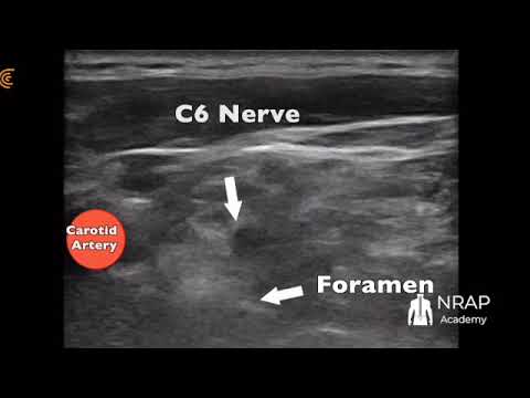
Ultrasound-guided technique. Ultrasound guidance for cervical selective nerve root injections (most commonly at C6 and C7, which are relatively superficial and have reliably identifiable sonoanatomy) allows real-time visualization of the nerve root, the surrounding vasculature (including radicular and vertebral vessels) with color Doppler, and the needle trajectory, which can reduce the risk of vascular puncture compared to a purely fluoroscopic approach. The C6 transverse process has a prominent anterior tubercle (Chassaignac's tubercle) that serves as a key sonographic landmark; the nerve root is typically identified as a hypoechoic oval structure within the trough between the anterior and posterior tubercles of the transverse process, often with the vertebral artery visible just anterior and medial. Color Doppler is used before and during needle advancement to actively avoid the vertebral artery and smaller radicular feeder vessels. Many practitioners use ultrasound and fluoroscopy together (ultrasound for real-time soft tissue and vascular visualization, fluoroscopy with contrast for confirmation of needle position and to rule out intravascular or intrathecal spread) to combine the strengths of both modalities. Because board questions frequently test "why do we use ultrasound here," the answer centers on direct visualization of vulnerable vascular structures that are invisible on fluoroscopy alone.
Interlaminar versus transforaminal approach. A cervical interlaminar epidural steroid injection places the needle into the posterior epidural space in the midline or paramedian plane, typically at C7–T1 or C6–C7, using a loss-of-resistance technique confirmed with fluoroscopic contrast. This approach treats multilevel or bilateral pathology and avoids the foraminal vasculature entirely, but it delivers medication less selectively to a single nerve root than a transforaminal approach. A transforaminal/selective nerve root approach delivers medication directly at the symptomatic foramen, which is more targeted both diagnostically and therapeutically, but carries the higher vascular risk discussed below because the needle passes near the vertebral artery and radicular arteries that can supply the anterior spinal artery and, in rare cases, the spinal cord itself.
Key Complications and Board Pitfalls
Complications of cervical transforaminal injections are a recurring board topic precisely because the cervical spine's vascular anatomy makes this one of the highest-risk common interventional pain procedures.
- Vertebral artery injury or injection. The vertebral artery runs through the transverse foramina of C6 through C1 and is directly adjacent to the exit path of the cervical nerve roots. Inadvertent intra-arterial injection of particulate steroid into the vertebral artery (or into a radicular artery that communicates with it) can cause embolic infarction of the brainstem or cerebellum, even with a very small injected volume, because these arteries supply critical posterior circulation territory with little collateral flow.
- Spinal cord infarction from radicular artery injection. Some patients have a radicular artery (sometimes called an artery of Adamkiewicz-type feeder in the cervical spine) that contributes to the anterior spinal artery supplying the spinal cord. Particulate steroid injected into one of these feeder vessels can cause catastrophic, often permanent, anterior spinal cord infarction and paralysis. This is one of the main reasons many practitioners now preferentially use non-particulate steroids (such as dexamethasone) for cervical transforaminal injections, since non-particulate steroids do not aggregate and embolize vessels the way particulate steroids (such as triamcinolone or methylprednisolone) can.
- Intravascular injection without infarction. Even without causing stroke, intravascular uptake of local anesthetic can cause rapid-onset seizure or cardiovascular collapse due to direct vertebral arterial delivery to the brain in very small volumes — this is why real-time contrast visualization (ideally with digital subtraction) and a slow, incremental, test-dose injection technique with frequent aspiration are standard of care.
- Dural puncture and intrathecal or subdural injection can occur with either interlaminar or transforaminal approaches and can cause high spinal anesthesia, total spinal anesthesia, or post-dural-puncture headache depending on volume and location.
- Nerve root injury, infection, and epidural hematoma are standard injection risks shared with other spinal procedures and should be discussed as part of informed consent, along with the vascular risks above that are specific to the cervical region.
- Transient complications such as vasovagal response, increased pain, or temporary upper extremity weakness/numbness from local anesthetic spread to the brachial plexus or phrenic nerve (causing transient diaphragmatic paresis, which matters in patients with compromised pulmonary reserve) are more common and generally self-limited.
For board purposes, the single highest-yield complication concept is this: because particulate steroids can embolize and cause permanent neurologic injury if injected intravascularly near the vertebral artery or a radicular feeder vessel, many protocols favor a non-particulate steroid for cervical transforaminal injections, combined with live or digital subtraction fluoroscopic contrast confirmation (and increasingly ultrasound with color Doppler) before any injectate is delivered.
Natural History and Prognosis
Most patients with acute cervical radiculopathy improve substantially with conservative, non-surgical management over a period of weeks to a few months. Many disc herniations undergo at least partial resorption over time as the extruded material is resorbed, and the inflammatory response around the nerve root that drives much of the pain and sensitization also tends to subside as the acute process settles. This favorable natural history is a major reason why conservative management — activity modification, physical therapy, and oral medications — is appropriately tried first in patients who do not have red-flag findings (progressive motor deficit, myelopathy, suspected infection or malignancy) or rapidly progressive neurologic deficits.
It is worth noting that the different components of a cervical radiculopathy do not always resolve at the same rate: pain and sensory symptoms often improve before an associated motor deficit has fully recovered, and mild residual findings, such as a subtle reflex asymmetry, can persist even after a patient has returned to normal function.
A minority of patients do not follow this generally favorable course. Those with persistent or progressive symptoms that fail to respond adequately to an appropriate trial of conservative care, and in many cases to image-guided injections as well, are reasonable candidates for surgical evaluation. When surgery is truly indicated and the patient is well-selected — particularly when there is a clear structural correlate on MRI that matches the clinical presentation — outcomes after surgical decompression are generally good, with most well-selected patients experiencing meaningful improvement in pain and function. Patients with less clear-cut clinical-radiographic correlation, such as predominantly axial neck pain or multilevel degenerative changes without an obviously corresponding symptomatic level, tend to have less predictable surgical outcomes, which is part of why careful patient selection matters so much before a surgical referral is made.
When to Refer for Surgical Evaluation
Several clinical scenarios should prompt consideration of surgical referral in a patient with cervical radiculopathy. Progressive or severe motor weakness is one of the clearest indications, since nerve root compression that is actively causing worsening strength is less likely to improve with continued conservative management alone. Signs of myelopathy — hyperreflexia, a positive Hoffmann sign, gait disturbance, or bowel or bladder dysfunction — are similarly important, both because they suggest spinal cord involvement rather than an isolated nerve root problem and because bowel or bladder dysfunction is, on its own, a general red-flag finding that warrants urgent evaluation regardless of whether a cervical radiculopathy is also present.
Failure of an adequate trial of conservative management is another standard indication for referral. Most protocols consider roughly six to twelve weeks of physical therapy, activity modification, and appropriate oral medications, together with at least one image-guided injection in appropriate candidates, to be a reasonable threshold before concluding that non-surgical measures are unlikely to be sufficient on their own. Severe, function-limiting pain that significantly impairs a patient's quality of life despite this conservative trial is also a reasonable basis for referral, even in the absence of significant weakness or myelopathic signs.
Before a surgical referral is made, MRI correlating with the patient's clinical level and findings is essentially required, since surgical planning targets a specific structural lesion rather than a clinical syndrome alone. Common surgical options include anterior cervical discectomy and fusion (ACDF) and posterior cervical foraminotomy; the choice between these approaches depends on factors such as whether the pathology is central or foraminal, how many levels are involved, and surgeon and patient preference. Detailed surgical decision-making is beyond the scope of pain medicine practice, but understanding the general options is useful context when counseling patients about what a surgical referral may ultimately lead to.
Video: Ultrasound-Guided C6 Nerve Root Injection
The video below walks through an ultrasound-guided approach to the C6 nerve root, including identification of the anterior and posterior tubercles of the transverse process, the nerve root itself, and the surrounding vasculature with color Doppler before needle advancement.
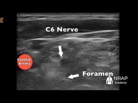
If the player above does not load, click the thumbnail to watch "Ultrasound Guided C6 Nerve Injection" on YouTube.
Keep Learning
For more on cervical epidural technique and the evidence behind it, listen to "Is Local Anesthetic in Cervical Epidurals Beneficial?" on The PainExam Podcast, where Dr. David Rosenblum reviews the evidence on local anesthetic dosing in cervical interlaminar epidural injections and discusses how ultrasound can complement fluoroscopic guidance for cervical radiculopathy. You can also find more ultrasound-guided spine procedure videos on the NRAP Academy YouTube channel. For related high-yield topics, see our posts on CRPS and the stellate ganglion block, PRP for chronic joint and spine pain, and caudal epidural injections for lumbar radiculopathy and spinal stenosis.
NRAP Academy Courses
If you're preparing for ABA Pain Medicine boards or want hands-on practice with ultrasound-guided spine and peripheral nerve procedures, NRAP Academy offers a Pain Management Board Review and Refresher Course and dedicated ultrasound training programs.
Looking for a Pain Specialist?
Looking for a pain specialist? Visit RosenblumPain.com or call (718) 436-7246. Patients outside the NYC/Brooklyn area can also book a virtual second-opinion consultation through RosenblumPain.com.
Disclaimer: This content is provided for general educational and informational purposes only and is intended for healthcare professionals, trainees, and patients seeking general information. It is not medical advice, does not constitute a diagnosis or treatment recommendation for any individual, and does not create a physician-patient relationship. Treatment decisions must always be individualized by a qualified, licensed healthcare provider based on a patient's specific history, examination, and current evidence-based guidelines. Dr. David Rosenblum, NRAP Academy, and PainExam make no warranties as to the completeness or applicability of this information and assume no liability for actions taken based on it. If you are a patient, please consult your own physician before making any medical decisions, and seek immediate medical attention for any urgent or emergency symptoms.