Ultrasound Guided Regional Anesthesia: A Clinical Guide
Introduction
Ultrasound-guided regional anesthesia (UGRA) has become a core skill in modern anesthesiology and pain management. By allowing direct visualization of nerves and surrounding structures, ultrasound significantly improves the precision and safety of regional anesthesia (RA). This technique has revolutionized the way nerve blocks are performed, reducing complications and improving patient comfort.
For physicians, anesthesiology residents, and pain specialists, understanding how to perform ultrasound-guided regional anesthesia is essential. Mastery of this technique not only enhances patient outcomes but also contributes to better workflow efficiency and a reduced reliance on systemic pain medication.
This clinical guide will walk you through the key steps involved in UGRA, from preparation and probe placement to needle guidance and anesthetic delivery. We’ll also discuss important safety considerations and offer insights into continuing medical education (CME) opportunities to help you grow your skills. Our goal is to provide a reader-friendly, evidence-based walkthrough with patient safety at its core.

Section 1: Understanding Ultrasound-Guided Regional Anesthesia
Ultrasound-guided regional anesthesia is a technique where ultrasound imaging is used to visualize nerves, blood vessels, and tissue planes before injecting local anesthetic near the targeted nerves. This method has largely replaced the traditional "blind" or landmark-based techniques due to its superior accuracy and reduced risk of complications.
UGRA is used in a wide range of settings, including orthopedic surgery, labor and delivery, outpatient procedures, and chronic pain management. It is also an excellent alternative for patients who are not ideal candidates for general anesthesia.
Common regional anesthesia techniques include:
- Brachial plexus blocks (interscalene, supraclavicular, infraclavicular)
- Femoral and sciatic nerve blocks
- Transversus abdominis plane (TAP) block
- Popliteal nerve block
- Fascia iliaca block
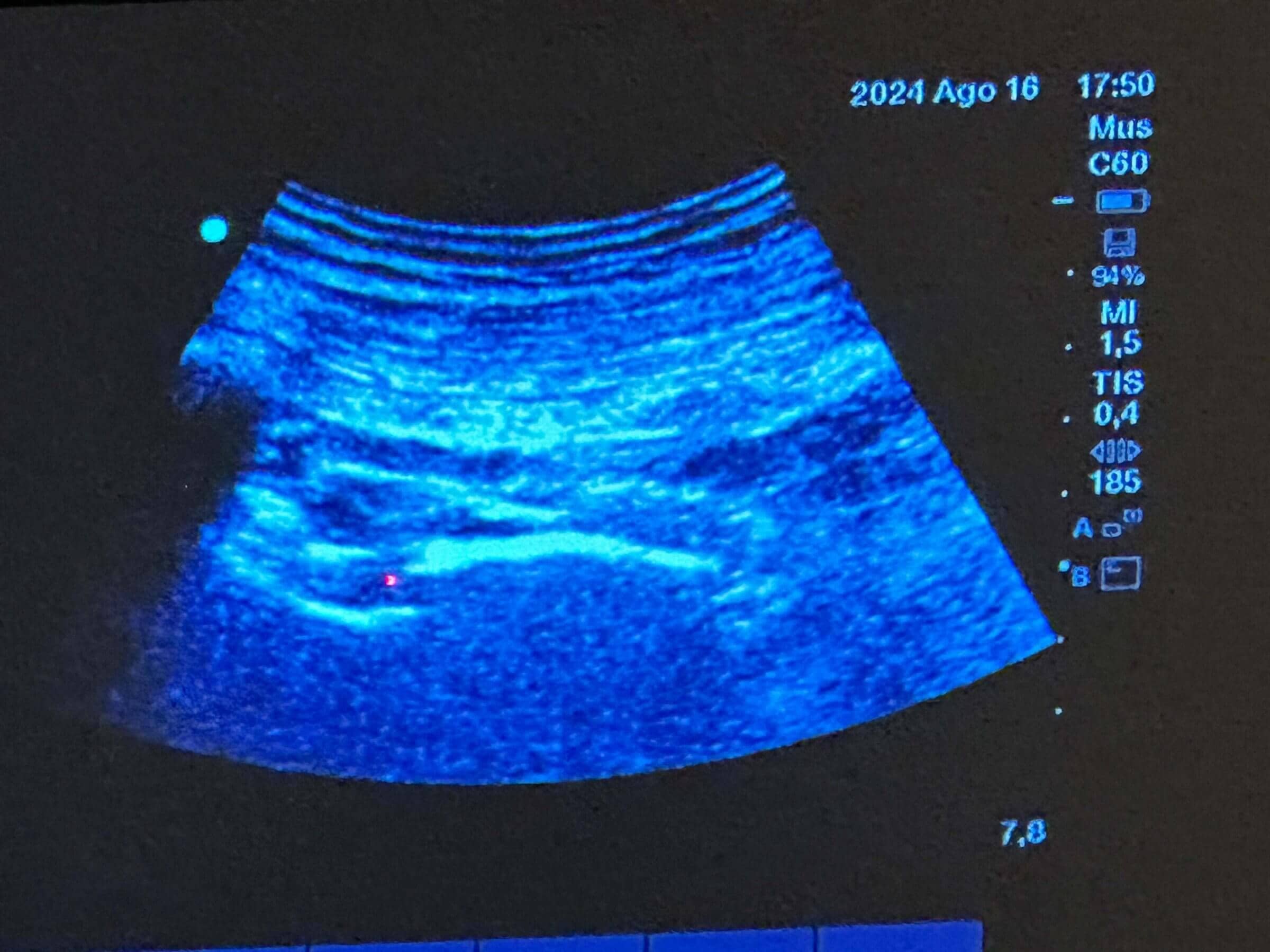
Ultrasound allows anesthesiologists to see the nerve, track the needle in real time, and observe the spread of local anesthetic. This level of precision helps minimize risks such as nerve injury, intravascular injection, and local anesthetic systemic toxicity (LAST). To enhance your understanding of sonographic views and structures, explore our Ultrasound Image Gallery.
Section 2: Preparing for the Procedure – A Complete RA Checklist
A successful regional block starts with careful planning. Following a structured checklist ensures both patient safety and procedural efficiency.
Patient Preparation
- Obtain informed consent, explaining the procedure, benefits, and risks.
- Review patient history for bleeding disorders, allergies, or previous adverse reactions.
- Ensure intravenous access and baseline vital signs are recorded.
Equipment Setup
- High-frequency linear probe (for superficial blocks) or low-frequency curvilinear probe (for deeper blocks)
- Sterile ultrasound probe cover and sterile gel
- Block needle (typically echogenic and 50–100 mm in length)
- Local anesthetic in appropriate volume and concentration
- Syringes, sterile gloves, drapes, and skin prep solution
Ultrasound Machine Settings
- Set the correct depth and gain for optimal image clarity.
- Use color Doppler to identify and avoid blood vessels.
- Freeze and store key images for documentation.
Sterile Technique
- Strict asepsis should be maintained throughout the procedure.
- Use sterile covers and drapes, and disinfect the skin thoroughly.
- Confirm proper hand hygiene and use of PPE.
Documentation Essentials
- Note the block type, side, drug used (with dose), patient response, and any complications.
- Record pre- and post-procedure assessments.
Thorough preparation is a core component of patient safety. To further your training, consider attending one of our Ultrasound Training Workshops designed for hands-on learning.
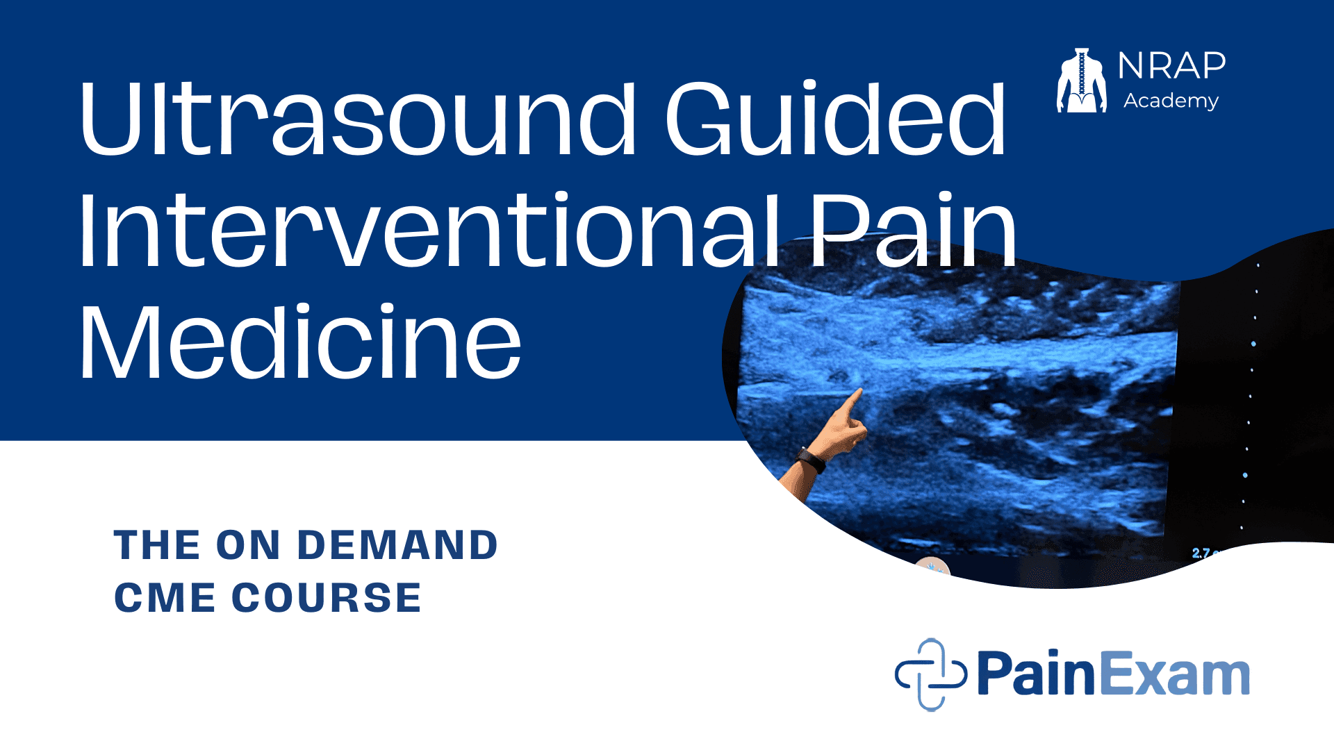
Section 3: Step-by-Step Guide on How to Perform Ultrasound-Guided Regional Anesthesia
Learning how to perform ultrasound-guided regional anesthesia requires both theoretical knowledge and hands-on practice. Below is a clinical walkthrough to guide you through a typical nerve block procedure:
Step 1: Choosing the Right Regional Anesthesia Technique
Each block serves a different surgical or diagnostic purpose. For example:
- Interscalene block: Shoulder surgery
- Femoral nerve block: Knee arthroplasty
- TAP block: Abdominal surgery
Identify the target nerve based on the planned procedure and patient anatomy.
Step 2: Anatomical Landmarks and Probe Placement
- Position the patient to expose the target area comfortably.
- Place the probe in the correct orientation. For example, for a supraclavicular brachial plexus block, position the probe just above the clavicle, perpendicular to the skin.
- Adjust the angle and pressure of the probe to enhance visibility.
- Use color Doppler to avoid vascular structures.
Step 3: Visualizing Nerve Structures
- Nerves appear as round or oval hyperechoic (bright) structures, often with a honeycomb pattern.
- Surrounding tissues like muscle (darker) and bone (white with shadowing) help with orientation.
Step 4: Needle Insertion and Real-Time Tracking
- Insert the needle either in-plane (along the probe’s length) or out-of-plane (perpendicular to the probe).
- In-plane insertion is preferred for better visualization of the entire needle.
- Advance slowly while watching the needle tip and adjusting the probe to keep it in view.
- Never inject unless the needle tip is clearly visible.
Step 5: Local Anesthetic Administration and Spread
- Aspirate to confirm you’re not in a vessel.
- Inject 1–2 mL of local anesthetic to confirm proper spread around the nerve.
- Continue injecting slowly while observing circumferential spread of anesthetic.
- Adjust needle position if spread is inadequate.
Tips to Avoid Complications
- Always visualize the full needle tip.
- Avoid injecting near vascular structures.
- Use minimal effective dose to reduce risk of systemic toxicity.
- Monitor the patient for 30 minutes post-procedure.
For deeper insights into block techniques and exam preparation, explore our Advanced Anesthesiology Board Review Course.
Section 4: Ultrasound Guided Regional Anesthesia Probe Placement Techniques
Effective probe handling is essential to UGRA success. Understanding how to optimize your image is just as important as needle technique.
Types of Probes
- Linear Probe (6–15 MHz): Ideal for superficial nerves such as the brachial plexus or femoral nerve.
- Curvilinear Probe (2–5 MHz): Suitable for deep structures like the sciatic nerve in larger patients.
Probe Movements
- Sliding: Move the probe across the skin to follow nerve paths.
- Tilting: Adjust the probe angle to improve reflection.
- Rotating: Align the probe to the anatomical plane of interest.
Needle Approach
- In-Plane: Provides continuous needle visualization.
- Out-of-Plane: Offers a more compact view but limited needle tip visibility.
Image Optimization Tips
- Adjust depth to keep nerves centered.
- Use adequate gel and apply consistent pressure.
- Re-scan after injection to confirm anesthetic spread.
A strong foundation in ultrasound physics and anatomy will support these technical skills. You can enhance your imaging knowledge by visiting NRAP Pain’s Homepage for expert resources and training options.
Section 5: Safety Considerations and Managing RA Complications in Ultrasound Guided Regional Anesthesia
Despite being safer than blind techniques, UGRA is not risk-free. Vigilance and adherence to protocols are essential.
Common Complications
- Nerve injury: Caused by direct trauma or high-pressure injection.
- Vascular puncture: May lead to hematoma or intravascular injection.
- Local anesthetic systemic toxicity (LAST): Rare but life-threatening.
Preventive Measures
- Always aspirate before injection.
- Monitor injection pressure; avoid resistance.
- Use ultrasound to confirm spread around the nerve, not into it.
- Limit local anesthetic volume according to patient weight.
Post-Block Monitoring
- Assess motor and sensory function.
- Monitor for signs of LAST (e.g., tinnitus, seizures, cardiac changes).
- Provide clear post-procedure instructions and emergency contact details.
Documentation is key to patient safety and legal protection. Maintain detailed records of every block performed. Want to solidify these protocols through CME? Check out our MOCA Anesthesia Board Review Course for in-depth content.
Section 6: Continuing Medical Education (CME) and Skill Advancement in Ultrasound Guided Regional Anesthesia
Mastery of UGRA is a journey, not a destination. With new techniques and devices emerging regularly, continuous learning is essential.
Why CME Matters
- Keeps you updated on the latest standards and technologies
- Enhances patient safety through evidence-based practice
- Helps fulfill licensing and credentialing requirements
Educational Formats
- Workshops and Simulations: Provide hands-on practice in a controlled environment
- Online CME Courses: Flexible, self-paced learning
- Certification Programs: Boost credibility and confidence
The National Regional Anesthesia & Pain Society (NRAP) offers a range of ultrasound training programs designed for physicians and residents. These include live courses, board review prep, and advanced CME workshops tailored to your career stage.
Ultrasound Guided Regional Anesthesia
Ultrasound-guided regional anesthesia is one of the most impactful advancements in modern anesthesia practice. It offers better accuracy, fewer complications, and improved patient outcomes. By mastering probe placement, image interpretation, and needle guidance, clinicians can perform safer, more effective nerve blocks.
Whether you’re a seasoned anesthesiologist or just beginning your training, continuous education and structured practice are the keys to excellence. Explore the clinical resources, CME opportunities, and training programs available through NRAP Pain to elevate your skills and confidence in regional anesthesia.
FAQs about Ultrasound Guided Regional Anesthesia
1. What are the most commonly used regional anesthesia techniques?
The most frequently used techniques include the brachial plexus block (interscalene, supraclavicular), femoral nerve block, sciatic nerve block, and TAP block. Each is chosen based on surgical location and patient needs.
2. How do I choose the right probe for a nerve block?
Use a linear probe (high frequency) for superficial nerves and a curvilinear probe (low frequency) for deeper structures. The probe choice depends on depth, body habitus, and target nerve.
3. What complications should I be aware of in RA?
Common complications include nerve damage, vascular puncture, infection, and local anesthetic systemic toxicity. Real-time ultrasound reduces these risks by improving accuracy.
4. Can residents perform ultrasound-guided RA safely?
Yes, with proper supervision and training, residents can perform UGRA effectively. Starting with simpler blocks and simulation training helps build foundational skills.
5. Where can I get CME training in regional anesthesia?
The NRAP offers CME-approved workshops, online board review courses, and ultrasound training designed for all experience levels. Visit the Ultrasound Training Page to learn more.