
Measure the patient, not just the needle. Accurate supraclavicular block depth measurement starts with ultrasound: locate the brachial plexus beside the subclavian artery and the corner pocket above the first rib, then measure the straight-down distance from skin to target before estimating the longer, angled path your needle will take. One study reported averages of 1.7 cm vertically and 3.7 cm along the planned path — neither is a universal insertion depth.
This article is intended for licensed clinicians and trainees who treat pain. Patients seeking personal care should visit RosenblumPain.com.
How to Measure Supraclavicular Block Depth

These two measurements help you choose a needle that can reach the target without treating its full length as a safe advancement limit. For structured hands-on instruction in ultrasound-guided procedures, see NRAP Academy’s Ultrasound Guided Interventional Pain Management Training. Keep the needle tip in view, identify the pleura, and stop if the tip or its path becomes unclear.
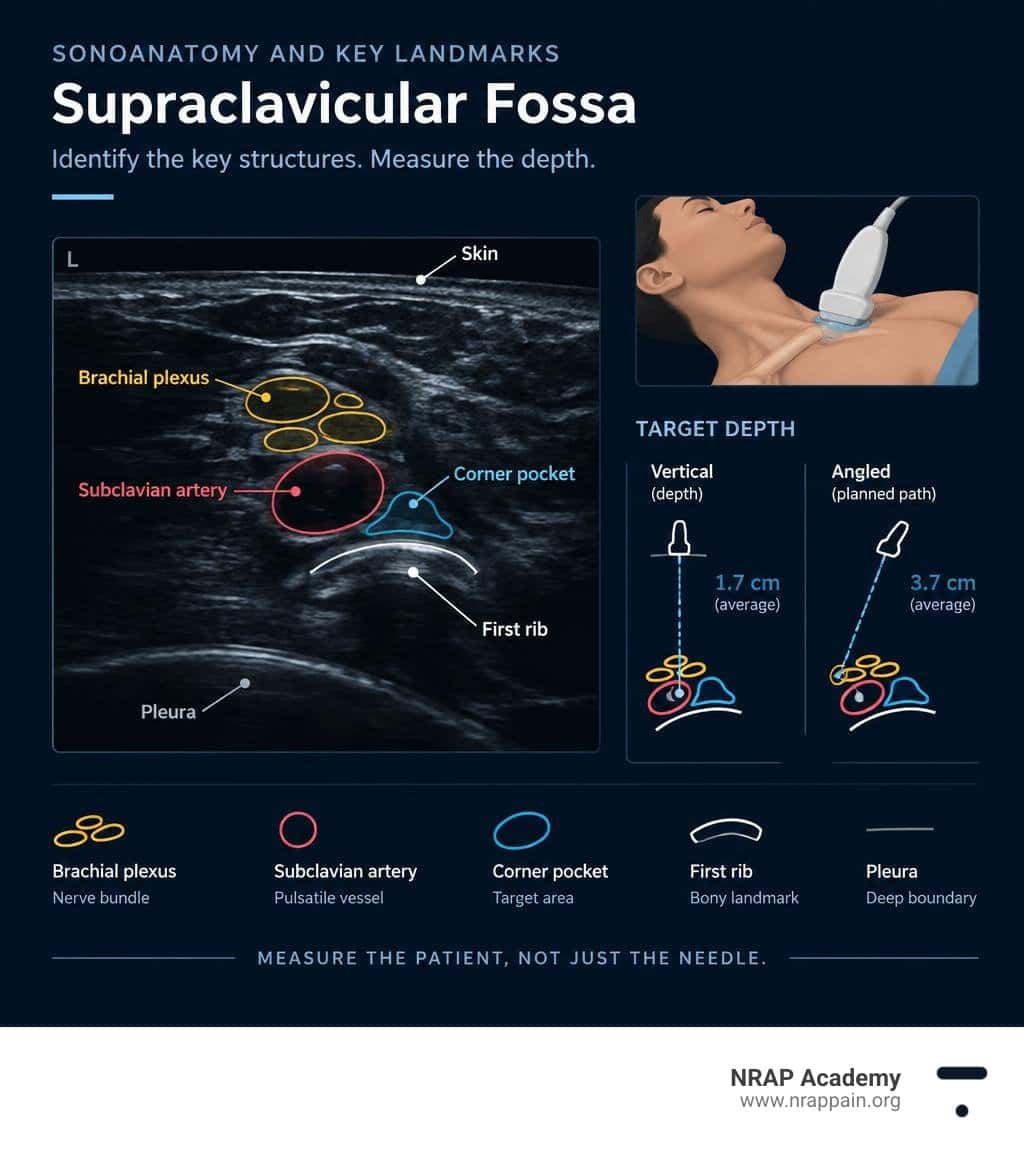
Sonoanatomy and Key Landmarks of the Supraclavicular Fossa
Performing a supraclavicular block has earned the affectionate nickname “the spinal of the upper extremity” because it provides dense, reliable anesthesia across the shoulder, arm, forearm, and hand. Because the neural trunks and divisions cross over the first rib in a compact bundle, a modest volume of local anesthetic achieves dense surgical anesthesia. However, this compact anatomy sits next to critical vascular and pulmonary structures.
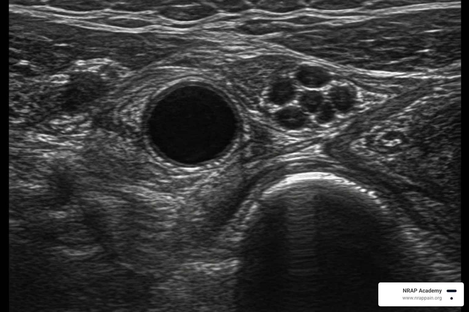
Understanding the acoustic landscape requires mastering key visual landmarks:
- Subclavian Artery (SA): A prominent, pulsatile, anechoic round structure resting directly on the bony floor of the supraclavicular fossa.
- Brachial Plexus (BP): A cluster of round or oval hypoechoic nodules with hyperechoic rims, often described as a “cluster of grapes” or a honeycomb pattern, located immediately superior and posterolateral to the subclavian artery.
- First Rib: A dense, hyperechoic curved line immediately deep to the subclavian artery and brachial plexus that casts a complete acoustic shadow beneath it.
- Pleura and Lung Dome: Located medial and deep to the first rib, the pleura appears as a shimmering, bright hyperechoic line that moves with respiration (“lung sliding”). An acoustic “comet-tail” artifact frequently projects below this boundary.
- Anterior and Middle Scalene Muscles: Muscular landmarks framing the trunks of the plexus as they exit the neck.
When evaluating these structures under ultrasound-guided regional anesthesia principles, you must maintain a clear view of the first rib. The bone serves as a physical acoustic shield: if your needle trajectory remains over the hyperechoic cortex of the first rib, you avoid accidental puncture of the deeper, medial pleural dome.
Sonographic Identification and Supraclavicular Block Depth Measurement in the Corner Pocket
To identify the corner pocket, place your linear transducer in the supraclavicular fossa posterior to the midpoint of the clavicle, directing the ultrasound beam caudally into the thoracic inlet.
Achieving an optimal acoustic view requires subtle probe adjustments:
- Tilting (Anterior/Posterior): Slight variations in probe tilt eliminate anisotropy, transforming dark, indistinct neural structures into crisp, honeycomb hypoechoic bundles.
- Centering: Ensure the intersection of the subclavian artery and the first rib is centered on your display monitor.
- Acoustic Shadow Verification: Confirm that the hyperechoic first rib sits directly underneath your target, casting a clear shadow.
Clinicians looking to hone their diagnostic image acquisition and probe alignment can build these scanning habits through Ultrasound Guided Interventional Pain Management Training.
Step-by-Step Guide to Supraclavicular Block Depth Measurement
Accurate calibration of tissue depth before needle entry eliminates procedural guesswork. Following a structured sonographic measurement protocol helps ensure clinical safety and efficacy during peripheral nerve blocks.
Pre-Scanning Protocol and Machine Optimization
- Transducer Selection: Use a high-frequency (10–15 MHz or 13–6 MHz) broadband linear array transducer. High frequencies provide the fine axial and lateral resolution needed to see individual nerve fascicles and the thin pleural line.
- Initial Depth Setting: Set your initial image depth to 4 to 6 cm. This wide view ensures you capture the subclavian artery, the first rib, and the pleural line on a single screen.
- Focal Zone Optimization: Place the ultrasound focal point directly at or slightly below the depth of the corner pocket (usually 1.5 to 2.5 cm).
- Gain and Time-Gain Compensation (TGC): Adjust the overall gain so the lumen of the subclavian artery is completely anechoic (black), while the fascial boundaries and neural epineurium are clearly hyperechoic (bright).
- Doppler Interrogation: Apply color Doppler briefly to detect any aberrant arterial vessels, such as the transverse cervical artery or dorsal scapular artery, crossing the planned needle path.
Measuring Shortest Distance (SD) and Longest Distance (LD)
Once you obtain a frozen image showing the plexus, artery, first rib, and pleura, use electronic ultrasound calipers to measure two distinct depths:
1. Shortest Distance (SD):
- Target: The vertical, 90-degree distance from the skin surface straight down to the superficial aspect of the brachial plexus or to the corner pocket floor.
- Clinical Value: In clinical trials, the mean shortest distance from skin to the most superficial neural element was 0.60 ± 0.262 cm, while the mean vertical depth to the deep corner pocket was 1.7 ± 0.8 cm (Yadav et al., 2016; Mistry et al., 2016).
- Application: Establishes tissue thickness over the neurovascular bundle and confirms how close the superficial nerves sit to the skin.
2. Longest Distance (LD):
- Target: The planned in-plane needle path measured from the lateral skin entry site diagonally to the corner pocket at the junction of the first rib and subclavian artery.
- Clinical Value: Studies report a mean in-plane needle trajectory distance of 3.7 ± 0.2 cm (with ranges spanning 2.5 to 5.2 cm depending on body habitus).
- Application: Determines the required length of your needle shaft and establishes an absolute advancement limit.
Clinical Predictors and Demographic Variations in Plexus Depth
Target depth varies widely across patient populations. Relying on average anatomical distances without individualized measurement increases the risk of under- or over-advancement.
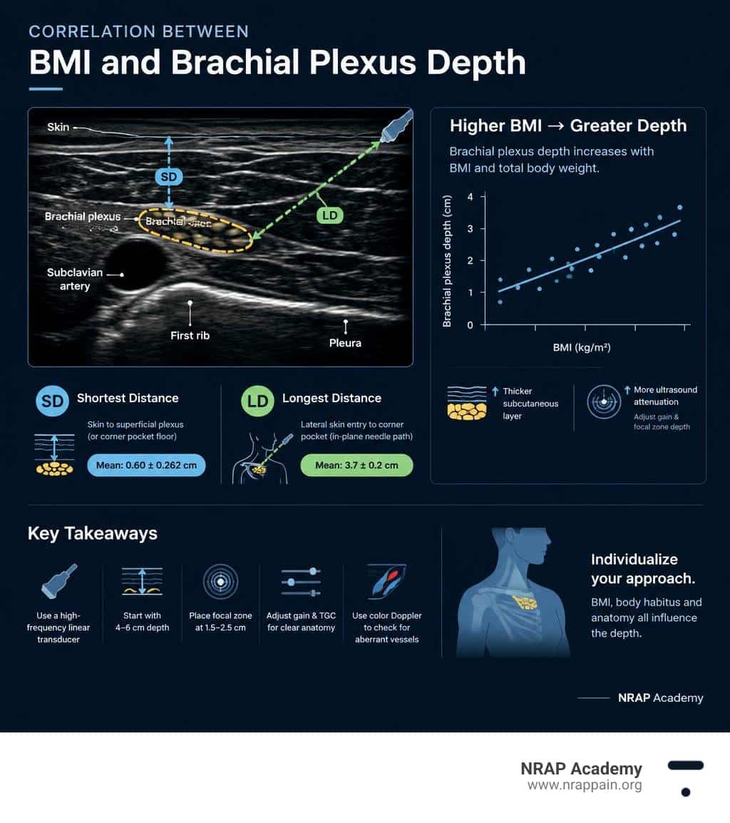
Impact of Patient BMI on Supraclavicular Block Depth Measurement
Body mass index (BMI) and total body weight correlate positively with brachial plexus depth (Yadav et al., 2016; Mistry et al., 2016). As adipose tissue accumulates in the supraclavicular fossa:
- The subcutaneous layer thickens, increasing the vertical skin-to-plexus distance.
- Ultrasound beam attenuation increases, requiring adjustments in gain and focal zone depth.
- The needle entry point must shift more laterally to maintain a shallow insertion angle (<30–45 degrees), which lengthens the overall needle path (LD).
Conversely, patient age and height show no strong correlation with corner pocket depth.
Special Population Adjustments: Pediatric to Obese Patients
| Patient Cohort | Typical Vertical Depth (SD) | Typical In-Plane Trajectory (LD) | Transducer Selection | Recommended Needle Length |
|---|---|---|---|---|
| Pediatric (< 30 kg) | 0.4–1.0 cm | 1.2–2.5 cm | High-frequency Linear (13–18 MHz, small footprint) | 25–35 mm (22–25G) |
| Normal Adult (BMI 19–25) | 1.0–1.8 cm | 2.8–3.8 cm | High-frequency Linear (10–15 MHz, standard 38 mm) | 38–50 mm (22G) |
| Obese Adult (BMI > 30) | 1.8–3.2 cm | 4.0–6.5 cm | Mid-to-low Linear (10–5 MHz) or Curved Array | 50–80 mm (21–22G) |
- Pediatric Patients: The plexus sits superficially, often less than 1 cm beneath the skin. Small needle movements produce large relative changes in position, making continuous tip tracking essential.
- Patients with Obesity: When adipose tissue pushes target structures past 3.5 to 4.0 cm deep, high-frequency linear probes may lose resolution. Switching to a lower-frequency probe (e.g., 8–5 MHz) maintains visibility of the first rib and pleura.
Needle Selection and Insertion Limits for Patient Safety
Safe needle advancement requires keeping the tip visible throughout its entire trajectory. Most regional anesthesia safety guidelines, including the European Society of Anaesthesiology recommendations, emphasize using an in-plane approach and confirming the needle tip location before injection.
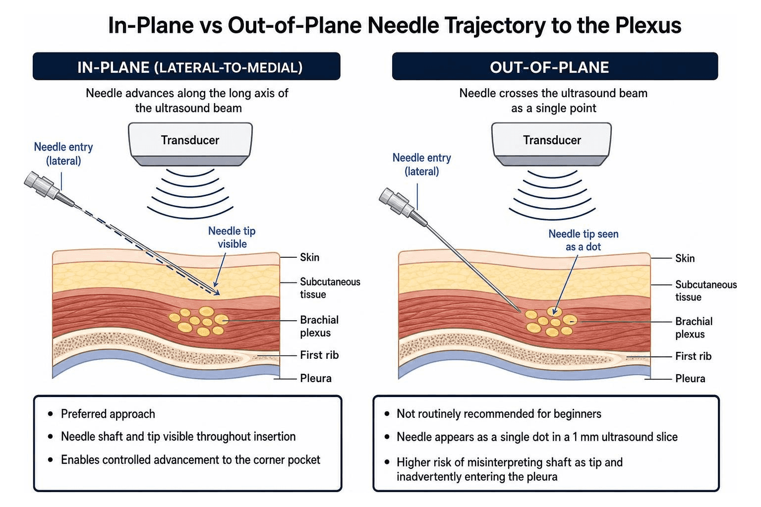
- In-Plane (Lateral-to-Medial): The preferred approach. The needle enters the skin on the lateral end of the transducer and advances along the long axis of the sound beam toward the corner pocket. This keeps the shaft and tip visible throughout insertion.
- Out-of-Plane: Not routinely recommended for beginners in the supraclavicular fossa because the needle crosses a narrow (1 mm) ultrasound slice as a single echogenic dot, making it easy to mistake the needle shaft for the tip and inadvertently enter the pleura.
Evaluating the 3 cm Needle Strategy vs Individualized Lengths
In average-weight adults, the deep neural elements sit at a mean depth of 1.34 ± 0.385 cm, and the corner pocket lies roughly 1.7 cm vertically below the skin (Yadav et al., 2016; Mistry et al., 2016). Consequently, a needle with a shaft length of 3 cm (30 mm) to 5 cm (50 mm) reaches the plexus sheath in most patients.
Using a standardized 3 cm needle offers a mechanical safety stop in lean individuals, preventing deep over-insertion toward the cupola of the lung. However, because the in-plane trajectory (LD) averages 3.7 cm, steep needle angles or higher BMIs often require a 50 mm needle.
A reliable safety rule is to never advance your needle deeper than the measured longest distance (LD) calculated during your pre-scan. Clinicians can refine their needle control through advanced interventional ultrasound education.
Mitigating Pneumothorax and Vascular Puncture Risks
Precise depth measurement and trajectory planning protect against common regional anesthesia complications:
- Pneumothorax Prevention: Always keep the hyperechoic first rib beneath your needle tip as a bony backstop. Never advance the needle beyond your measured LD or medial to the subclavian artery wall.
- Vascular Puncture & Toxicity: Aspirate every 3 to 5 mL during injection. Depositing local anesthetic slowly avoids systemic toxicity and vascular trauma.
- Phrenic Nerve Palsy: Ipsilateral phrenic nerve block occurs in approximately 50% of standard-volume supraclavicular blocks due to cephalad spread toward the anterior scalene muscle. Using ultrasound guidance allows you to reduce injectate volumes (e.g., 15–20 mL) while maintaining a dense block and lowering the risk of hemidiaphragmatic weakness.
- Adjuvant Safety: Adding evidence-based adjuvants such as dexamethasone or dexmedetomidine, as studied in clinical trials for upper limb nerve blockade, prolongs block duration without requiring higher volumes of local anesthetic.
- Lymph Node Differentiation: Pathological or reactive lymph nodes in the supraclavicular fossa can resemble neural elements. Distinguishing hypoechoic lymph nodes from nerve fascicles prevents direct nodal puncture, consistent with clinical criteria for assessing supraclavicular lymph nodes.
Conclusion
At NRAP Academy, we believe safe interventional practice starts with measurable anatomical precision. Integrating structured supraclavicular block depth measurement into your routine scanning protocol eliminates guesswork, guides appropriate needle selection, and protects critical structures like the subclavian artery and pleura.
For a procedure-specific educational resource, see NRAP Academy’s supraclavicular brachial plexus block training. For further clinician education in ultrasound-guided procedures, explore NRAP Academy’s Ultrasound Guided Interventional Pain Management Training.
Scientific References
- Yadav N, Ayub A, Garg R, Nanda S, Gupta B, Sawhney C. “Sonographic assessment of predictors of depth of the corner pocket for ultrasound-guided supraclavicular brachial plexus block.” Journal of Anaesthesiology, Clinical Pharmacology. 2016. PMCID: PMC4784208.
- Mistry T, Mangal V, Sharma G, Agrawal A. “Assessment of variation in depth of brachial plexus using ultrasound for supraclavicular brachial plexus block in patients undergoing elective upper limb surgery.” Indian Journal of Anaesthesia. 2016. PMCID: PMC4910478.
Medical Disclaimer
This article is intended exclusively for educational and examination review purposes. It does not constitute medical advice, does not establish a physician-patient relationship, and must not serve as the basis for clinical decision-making or patient care. Clinical standards, diagnostic guidelines, and pharmacotherapy regimens change frequently. Readers are solely responsible for independently verifying all clinical data, drug dosages, contraindications, procedural steps, and safety profiles against current primary literature, official manufacturer package inserts, and institution-specific clinical policies before administering any treatment. Neither the author nor NRAP Academy assumes any liability for injury, damages, or adverse outcomes resulting from the application of information contained in this article.


