Why Ultrasound Guidance Is Transforming Interventional Pain Practice

Interventional pain medicine has evolved significantly over the past two decades. New technologies have improved the precision of diagnostic and therapeutic procedures, allowing physicians to treat pain more effectively while minimizing risk.
One of the most important developments in this field is the use of ultrasound guided pain procedures. Ultrasound imaging enables physicians to visualize anatomical structures in real time, making procedures safer and more accurate.
Many physicians now incorporate ultrasound guidance into their daily practice after completing specialized training such as an anesthesia ultrasound course or an ultrasound nerve block course. These educational programs teach clinicians how to identify nerves, muscles, and joints while guiding needles with high precision.
As evidence supporting ultrasound guidance continues to grow, it is becoming a central component of modern interventional pain practice.

The Evolution of Imaging in Pain Medicine: Ultrasound Guided Pain Procedures
Medical imaging has always played a critical role in pain management procedures. Imaging techniques help physicians locate anatomical structures and ensure that injections are performed accurately.
Over time, new technologies have improved the safety and effectiveness of these procedures.
Traditional Imaging Methods
For many years, fluoroscopy has been the primary imaging modality used in interventional pain medicine. Fluoroscopy provides real-time X-ray images that allow physicians to guide needles during procedures such as epidural injections or spinal interventions.
While fluoroscopy remains useful in many cases, it also has certain limitations. It exposes both patients and physicians to ionizing radiation and may not clearly visualize soft tissues such as nerves or blood vessels.
These limitations have encouraged clinicians to explore alternative imaging techniques.
Emergence of Ultrasound Technology
Ultrasound imaging has become increasingly popular in pain medicine due to its ability to visualize soft tissues in real time. Unlike fluoroscopy, ultrasound uses sound waves rather than radiation.
This technology allows physicians to see muscles, tendons, nerves, and blood vessels during procedures. As a result, clinicians can perform injections with greater precision and improved safety.
Many physicians now incorporate ultrasound training into their professional development through pain medicine courses or specialized workshops.

What Are Ultrasound Guided Pain Procedures?
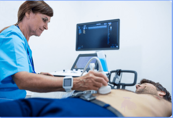
Ultrasound guided procedures involve using ultrasound imaging to guide needles during injections or nerve blocks. The physician holds an ultrasound probe on the patient’s skin while watching live images on a monitor.
This technique provides direct visualization of anatomical structures, making it easier to avoid sensitive tissues and place medication exactly where it is needed.
Basic Principles of Ultrasound Guidance
Ultrasound guidance relies on high-frequency sound waves that create detailed images of internal structures. When these waves interact with tissues, they reflect back to the probe and generate real-time images.
This method allows physicians to track the needle as it moves through tissue, ensuring accurate placement. Many anesthesia ultrasound course programs teach these techniques through hands-on training sessions.
Key principles of ultrasound-guided procedures include:
- identifying anatomical landmarks
- maintaining continuous visualization of the needle
- avoiding blood vessels and sensitive structures
- confirming proper medication placement
These skills are essential for performing safe and effective pain interventions.
Common Pain Procedures Performed with Ultrasound
Ultrasound guidance is now used for a wide range of procedures in pain medicine.
Some of the most common ultrasound-guided procedures include:
- peripheral nerve blocks
- musculoskeletal injections
- trigger point injections
- joint injections
- regenerative medicine injections
Physicians often refine these techniques through joint injection CME courses or advanced pain management course programs.

Key Benefits of Ultrasound in Pain Medicine
The growing popularity of ultrasound guidance is largely due to its many clinical advantages. Physicians who adopt this technology often report improved procedural accuracy and better patient outcomes.
These advantages have made ultrasound a valuable tool in modern interventional pain practice.
Real-Time Visualization
One of the greatest benefits of ultrasound in pain medicine is real-time visualization of anatomical structures. Physicians can observe the needle as it moves through tissue, allowing them to adjust the trajectory if necessary.
This dynamic imaging capability is particularly useful when performing nerve blocks or musculoskeletal injections.
Real-time visualization allows physicians to:
- identify nerves and blood vessels
- monitor needle placement continuously
- confirm proper medication spread
- reduce procedural complications
These benefits contribute to more precise and effective treatments.
Improved Safety and Accuracy
Ultrasound guidance also improves safety by allowing clinicians to avoid important structures such as blood vessels or organs. By visualizing these structures directly, physicians can minimize the risk of complications.
Training programs such as an ultrasound nerve block course help physicians develop the technical skills needed to perform these procedures safely.
As a result, ultrasound guidance is becoming the preferred method for many peripheral nerve and joint injections.

Radiation Reduction in Pain Medicine
Radiation exposure is a significant concern in interventional pain medicine. Procedures performed with fluoroscopy expose both patients and physicians to ionizing radiation.
Over time, repeated exposure can pose health risks.
Limitations of Fluoroscopy
Fluoroscopy provides valuable imaging for certain procedures, particularly spinal interventions. However, it also has drawbacks that have encouraged physicians to explore alternative imaging methods.
Some limitations include:
- exposure to ionizing radiation
- limited visualization of soft tissues
- requirement for protective equipment
These challenges have led many clinicians to incorporate ultrasound guidance into their practice.
Safer Imaging for Physicians and Patients
One of the most important advantages of ultrasound is the ability to perform procedures without radiation. This represents a major step forward in radiation reduction pain medicine.
By eliminating radiation exposure, ultrasound-guided procedures provide a safer environment for both physicians and patients. This benefit is particularly important for clinicians who perform large numbers of injections each year.

Fluoroscopy vs Ultrasound Pain Procedures
Understanding the differences between these imaging techniques helps physicians choose the most appropriate approach for each procedure.
Both methods have advantages, and many clinicians use them in complementary ways.
Differences in Imaging Techniques
Fluoroscopy and ultrasound rely on different technologies to produce images.
Fluoroscopy uses X-ray imaging, which is effective for visualizing bones and spinal structures. Ultrasound, on the other hand, excels at imaging soft tissues such as muscles, ligaments, and nerves.
Because of these differences, ultrasound is particularly useful for peripheral nerve blocks and musculoskeletal procedures.
Choosing the Right Modality
In many cases, the decision between fluoroscopy and ultrasound depends on the specific procedure being performed.
Some physicians choose to combine both technologies when appropriate. For example, ultrasound may be used to guide needle placement while fluoroscopy confirms final positioning.
Learning when to use each modality is an important component of advanced pain medicine courses.

Training Requirements for Ultrasound Guided Pain Procedures
Although ultrasound offers many advantages, physicians must receive proper training before incorporating it into their clinical practice.
Structured training programs help clinicians develop both theoretical knowledge and practical skills.
Importance of Formal Ultrasound Education
Formal education programs teach physicians how to interpret ultrasound images and perform guided procedures safely.
Many clinicians begin with an anesthesia ultrasound course, which introduces the fundamentals of ultrasound physics and anatomy.
These courses often include lectures, demonstrations, and hands-on practice.
Hands-On Clinical Training
Hands-on experience is essential for mastering ultrasound-guided procedures. Physicians must learn how to coordinate probe positioning, needle movement, and image interpretation simultaneously.
Training programs such as joint injection CME workshops provide opportunities for physicians to practice these skills in supervised environments.

Core Procedures Physicians Learn in Ultrasound Training
Physicians participating in ultrasound training programs learn a variety of procedures that are commonly used in pain management.
These techniques can significantly expand a clinician’s treatment options.
Ultrasound Nerve Blocks
Peripheral nerve blocks are among the most common ultrasound-guided procedures. These injections deliver anesthetic medication near specific nerves to block pain signals.
Physicians often learn these techniques during an ultrasound nerve block course, where they practice identifying nerves and guiding needles safely.
Joint Injection Techniques
Joint injections are another important component of pain management. These procedures can help relieve pain caused by arthritis or musculoskeletal injuries.
Clinicians frequently attend joint injection CME courses or complete an online joint injection course to improve their technique and learn new approaches.

Expanding Skills Through Pain Medicine Courses
Continuing education is essential for physicians who want to stay current with advances in pain medicine.
Participating in specialized training programs allows clinicians to refine their skills and learn emerging techniques.
CME Workshops and Clinical Training
Many physicians attend pain management course workshops that focus on practical procedural skills. These programs typically combine lectures with hands-on training sessions.
Workshops provide opportunities to practice injections, nerve blocks, and other interventional techniques under expert supervision.
Online and Hybrid Learning Options
Modern education programs often combine online learning with in-person training. Anesthesia courses online allow physicians to study theoretical material before attending hands-on workshops.
This hybrid model makes advanced training more accessible for busy clinicians.

Future of Ultrasound in Interventional Pain Medicine
Ultrasound technology continues to evolve, and its role in pain medicine is expected to expand further in the coming years.
New imaging techniques and treatment strategies are transforming the field.
Integration with Regenerative Medicine
Ultrasound guidance plays an important role in regenerative therapies. Procedures such as platelet-rich plasma injections require precise placement of biological materials.
Many physicians learn these techniques through specialized regenerative medicine courses.
Advanced Imaging and Precision Medicine
As imaging technology improves, ultrasound may become even more precise and versatile. New software tools and high-resolution probes are making it easier to visualize complex anatomical structures.
These advancements will continue to enhance the safety and effectiveness of ultrasound guided pain procedures.

Conclusion
Ultrasound technology is rapidly transforming the field of interventional pain medicine. By providing real-time visualization and eliminating radiation exposure, ultrasound guidance offers significant advantages over traditional imaging methods.
Physicians who complete advanced training programs such as pain medicine courses, joint injection CME courses, or an anesthesia ultrasound course can integrate these techniques into their clinical practice.
As more clinicians adopt ultrasound-guided techniques, patients will benefit from safer procedures, improved accuracy, and better pain relief outcomes.

FAQs
What are ultrasound guided pain procedures?
Ultrasound guided pain procedures involve using ultrasound imaging to visualize anatomical structures and guide needles during injections or nerve blocks.
Why is ultrasound preferred in many pain procedures?
Ultrasound allows real-time visualization of nerves, muscles, and blood vessels while eliminating radiation exposure.
What training is required for ultrasound guided pain procedures?
Physicians often complete programs such as an anesthesia ultrasound course, pain management course, or joint injection CME courses to develop these skills.
Can joint injections be learned online?
Yes. Many clinicians begin with an online joint injection course before attending hands-on workshops.
Are ultrasound techniques important for pain medicine certification?
Yes. Knowledge of ultrasound-guided procedures can support professional development and training pathways related to PM&R Board Certification and advanced pain medicine practice.