Table of Contents
- What Is Occipital Neuralgia? Anatomy of the Greater, Lesser, and Third Occipital Nerves
- How Occipital Neuralgia Is Diagnosed
- Occipital Neuralgia Treatment: From Conservative Care to Nerve Block
- Epidemiology and Prognosis
- Video: Ultrasound-Guided Lesser Occipital Nerve Block
- Keep Learning
- NRAP Academy Courses
- Looking for a Pain Specialist?
- FAQs
Occipital neuralgia is a common but frequently misdiagnosed cause of posterior headache, often mistaken for migraine, tension-type headache, or cervicogenic pain. For patients with sharp, shock-like pain over the back of the scalp, a carefully performed diagnostic nerve block can be both clarifying and therapeutic. This occipital neuralgia treatment guide reviews the anatomy, clinical diagnosis, and treatment ladder, with a focus on the ultrasound-guided lesser occipital nerve block technique.
In this post:
- What Is Occipital Neuralgia? Anatomy of the Greater, Lesser, and Third Occipital Nerves
- How Occipital Neuralgia Is Diagnosed
- Occipital Neuralgia Treatment: From Conservative Care to Nerve Block
- Epidemiology and Prognosis
- Video: Ultrasound-Guided Lesser Occipital Nerve Block
- Keep Learning
- NRAP Academy Courses
- Looking for a Pain Specialist?
What Is Occipital Neuralgia? Anatomy of the Greater, Lesser, and Third Occipital Nerves
Occipital neuralgia is a neuropathic pain syndrome defined by paroxysmal, shooting or stabbing pain in the distribution of the greater occipital nerve (GON), lesser occipital nerve (LON), and/or third occipital nerve (TON). The International Classification of Headache Disorders (ICHD-3) defines it as unilateral or bilateral pain in the posterior scalp, often with tenderness over the affected nerve and occasional dysesthesia or allodynia of the overlying skin. Patients frequently describe brief electric-shock-like jabs superimposed on a more constant dull ache, and many report that brushing their hair or resting the head on a pillow reproduces the pain.
All three occipital nerves are sensory branches derived from the dorsal rami of the upper cervical nerve roots, and understanding their relationships is essential for both diagnosis and image-guided treatment:
- Greater occipital nerve (GON) – arises primarily from the dorsal ramus of C2, with contributions from C3. It pierces the semispinalis capitis and trapezius (or the trapezial aponeurosis) roughly 2–3 cm lateral to the midline at the level of the inferior nuchal line, running alongside the occipital artery, which serves as a useful ultrasound landmark. It supplies sensation to the medial posterior scalp up to the vertex.
- Lesser occipital nerve (LON) – arises from the ventral rami of C2–C3 as part of the cervical plexus, not the dorsal rami. It ascends along the posterior border of the sternocleidomastoid (SCM) muscle and becomes subcutaneous in the upper neck/mastoid region before supplying the lateral posterior scalp and skin over the mastoid process. Because it travels superficially along the SCM’s posterior border, it is readily visualized with a high-frequency linear ultrasound transducer.
- Third occipital nerve (TON) – a superficial branch of the C3 dorsal ramus that crosses the C2-C3 facet joint before becoming cutaneous near the GON, supplying a small area of skin below the inferior nuchal line. It is clinically relevant in cervicogenic headache arising from the C2-C3 facet joint and is a common source of diagnostic confusion with occipital neuralgia.
For the lesser occipital nerve block specifically, the key anatomic relationships are the SCM muscle (the nerve runs just posterior to its posterior border), the mastoid process (a fixed bony landmark), and the investing layer of the deep cervical fascia, beneath which the nerve travels before piercing more superficially. Because the LON is a cervical plexus branch rather than a dorsal ramus branch like the GON, its course and depth are more variable between patients, which is precisely why ultrasound guidance offers a meaningful advantage over blind, landmark-based injection: it allows direct visualization of the nerve (or its expected fascial plane) alongside the external jugular vein and SCM, reducing the chance of intravascular injection or a missed target.
How Occipital Neuralgia Is Diagnosed
Occipital neuralgia is a clinical diagnosis supported by history, physical exam, and response to diagnostic nerve blockade. There is no definitive imaging test that confirms the diagnosis, but imaging plays an essential role in excluding secondary causes.
Typical clinical features include:
- Paroxysmal, shock-like or stabbing pain lasting seconds to minutes, in the distribution of the occipital nerves, often with a background dull ache between paroxysms.
- Pain that may radiate forward toward the vertex, temple, or behind the eye, which can mimic migraine or cluster headache.
- Point tenderness over the nerve’s exit point (the “Tinel-like” sign at the nuchal ridge for the GON, or over the mastoid/posterior SCM border for the LON).
- Allodynia of the scalp, with patients reporting pain from light touch, hair brushing, or wearing a hat.
- Possible temporary relief with pressure over the nerve or with a diagnostic local anesthetic block — a positive block is supportive of, though not solely diagnostic for, occipital neuralgia.
Red flags that warrant further workup before attributing posterior headache to occipital neuralgia include sudden “thunderclap” onset, new headache after age 50, fever or neck stiffness, focal neurologic deficits, papilledema, headache triggered by cough/Valsalva, a history of trauma, cancer, or anticoagulation, and progressive or treatment-refractory symptoms. These findings should prompt neuroimaging (MRI brain/cervical spine) to rule out structural causes such as Chiari malformation, posterior fossa lesions, cervical spine pathology, or vascular causes before proceeding with interventional treatment.
Because several conditions overlap with occipital neuralgia in presentation, a structured differential is useful:
| Condition | Key Distinguishing Features | Helpful Clues |
|---|---|---|
| Cervicogenic headache | Unilateral pain originating from the neck (often C2-C3 facet or disc), referred to the head; worsened by neck movement or sustained posture | Restricted cervical range of motion; relief with C2-C3 facet or medial branch (third occipital nerve) block |
| Migraine | Throbbing, often unilateral, associated with nausea, photophobia, phonophobia; may have aura | Episodic with migraine-specific triggers; less focal tenderness over occipital nerve exit points |
| Cluster headache / other TACs | Severe unilateral periorbital pain with autonomic features (lacrimation, ptosis, rhinorrhea) | Strict circadian/circannual periodicity; occipital nerve block may still provide adjunctive relief but anatomy of pain differs |
| Tension-type headache | Bilateral, band-like, pressure quality; not paroxysmal or shock-like | No focal nerve tenderness; associated with pericranial muscle tightness |
| Occipital neuralgia | Paroxysmal, shooting/electric pain in GON/LON/TON distribution with point tenderness and scalp allodynia | Positive diagnostic response to occipital nerve block; tenderness reproduces typical pain |
| Secondary structural causes (Chiari, mass, vascular) | Red-flag features; may have additional neurologic findings | Abnormal neuroimaging; progressive or atypical course |
Occipital Neuralgia Treatment: From Conservative Care to Nerve Block
Management of occipital neuralgia generally follows a stepwise escalation, moving from conservative and pharmacologic measures to image-guided interventions, and finally to more invasive neuromodulatory options for refractory cases.
Conservative measures
As part of occipital neuralgia treatment, first-line conservative measures include patient education, activity and posture modification, heat or ice application to the occiput, gentle cervical range-of-motion and stretching exercises, and physical therapy targeting the upper cervical and suboccipital musculature. Trigger point release and myofascial work over the suboccipital region can meaningfully reduce mechanical irritation of the nerves in patients whose symptoms are aggravated by muscle tension.
Medications for Occipital Neuralgia Treatment
Pharmacologic options for occipital neuralgia treatment are generally borrowed from the broader neuropathic pain and headache treatment literature, and are typically used as adjuncts rather than standalone cures:
- Anticonvulsants (e.g., gabapentinoids, carbamazepine-class agents) – often first-line for neuropathic, shooting pain qualities.
- Tricyclic antidepressants or SNRIs – useful for the chronic, dull background pain component and for comorbid tension-type or migraine features.
- Muscle relaxants – may help when cervical/suboccipital muscle spasm is a prominent contributor.
- NSAIDs – for acute flares, used judiciously given limited long-term benefit in neuropathic pain.
- Topical agents (lidocaine patches, topical NSAIDs) – low-risk options for localized tenderness over the nerve.
Medications are typically combined with conservative measures before moving to interventional treatment, particularly in patients with mild or infrequent symptoms.
Ultrasound-guided occipital nerve block
For patients with persistent or diagnostically ambiguous symptoms, an ultrasound-guided nerve block is a core component of occipital neuralgia treatment, serving as both a valuable diagnostic test and an effective therapeutic option. Compared with blind, landmark-based injection, ultrasound allows the proceduralist to directly visualize the nerve or its expected anatomic plane along with adjacent vascular structures (the occipital artery for the GON, and the external jugular vein and SCM vasculature for the LON), improving accuracy and reducing the risk of intravascular injection.
Lesser occipital nerve block – technique overview:
- Positioning: The patient is seated or placed prone/lateral with the neck slightly flexed and rotated away from the side being treated, exposing the posterior border of the SCM and mastoid region.
- Transducer selection and setup: A high-frequency linear transducer (typically 10–18 MHz) is used given the superficial location of the nerve.
- Scanning: The probe is placed transversely at the level of the mastoid process, identifying the SCM muscle. The transducer is then slid posteriorly to the posterior border of the SCM, where the lesser occipital nerve is often visualized as a small, round-to-oval hyperechoic structure in the subcutaneous tissue or just deep to it, typically accompanied by small vascular branches. When the nerve itself is not clearly resolved, the injection targets the fascial plane along the posterior SCM border where the nerve is known to travel.
- Needle approach: Using an in-plane or out-of-plane technique (operator preference), a short, fine-gauge needle is advanced toward the nerve or target fascial plane under direct real-time visualization, with care taken to identify and avoid the external jugular vein and small superficial vessels in the field.
- Injection: After negative aspiration, a small volume of local anesthetic (often combined with a corticosteroid for a more sustained anti-inflammatory effect in appropriate candidates) is injected incrementally around the nerve, watching for anechoic spread confirming correct plane placement (hydrodissection) rather than intraneural or intravascular injection.
- Expected result: Most patients experience rapid-onset numbness in the distribution of the lesser occipital nerve (posterolateral scalp over the mastoid region) within minutes, with a significant reduction in their typical paroxysmal pain. This immediate response is itself diagnostically useful — a robust, reproducible response supports the occipital nerve as the pain generator.
The greater occipital nerve block follows an analogous ultrasound-guided approach, with the transducer placed over the nuchal ridge to identify the nerve adjacent to the occipital artery between the semispinalis capitis and trapezius, or more superficially once it has pierced the trapezial fascia.
| Technique Point | Pearl |
|---|---|
| Vascular avoidance | Use color Doppler to identify the occipital artery (GON) or external jugular/superficial vessels (LON) before needle insertion |
| Needle visualization | Keep the needle shaft in-plane with the beam throughout advancement; the superficial target depth makes steep, out-of-plane approaches harder to track accurately |
| Injectate spread | Watch for an expanding anechoic halo around the nerve/fascial plane as confirmation of correct needle tip position before injecting the full volume |
| Diagnostic value | A robust, reproducible pain reduction after the block supports the occipital nerve as the pain generator and helps distinguish occipital neuralgia from cervicogenic headache or migraine |
| Steroid use | Reserve corticosteroid addition for select patients given limited data on repeated steroid exposure near superficial scalp tissue; many diagnostic blocks use local anesthetic alone |
Complications
As part of occipital neuralgia treatment, ultrasound-guided nerve blocks are generally low-risk procedures. Potential complications include transient dizziness or vasovagal response, local site soreness or bruising, infection (rare, with sterile technique), intravascular injection (minimized with ultrasound and Doppler), and, rarely, temporary hair loss or skin atrophy at the injection site with repeated corticosteroid use. Systemic local anesthetic toxicity is exceedingly rare given the small injectate volumes used in this superficial block.
Epidemiology and Prognosis
Occipital neuralgia is relatively uncommon compared with migraine or tension-type headache, both of which affect a much larger share of the population. That said, it is likely underdiagnosed, since its hallmark paroxysmal, shock-like pain is frequently mistaken for one of these more common headache types, particularly when point tenderness over the occipital nerve exit points is not specifically assessed. It can occur at any age, but is seen more often in middle-aged and older adults. Accurate diagnosis is a prerequisite for effective occipital neuralgia treatment, since mislabeling the condition as migraine or tension-type headache can delay appropriate care.
Several factors are recognized as associations or contributors to the development of occipital neuralgia, including:
- A history of whiplash or other cervical trauma, which can irritate or scar the tissue surrounding the occipital nerves.
- Prolonged poor posture or sustained neck flexion, including the repetitive “tech neck” positioning associated with extended smartphone, tablet, or computer use.
- Chronic tension or tightness in the suboccipital musculature, which can mechanically irritate the nerves as they pierce the overlying muscle and fascia.
- Less commonly, frank entrapment of the nerve by surrounding fascial bands or a compressive structural lesion – part of why red-flag features on history or exam warrant neuroimaging before interventional treatment proceeds.
Overall, the prognosis following occipital neuralgia treatment is generally favorable. Many patients improve substantially with conservative measures alone or in combination with a diagnostic-therapeutic nerve block, and a strong, reproducible response to a block is itself considered a favorable prognostic sign, supporting the occipital nerve as the primary pain generator. That said, a subset of patients follow a more chronic, relapsing course and require repeat blocks, radiofrequency ablation, or neuromodulation to maintain adequate control.
Patient education about posture, ergonomics, and avoidance of common triggers – such as tight-fitting hats, prolonged neck flexion, and sleeping positions that compress the occiput – is a reasonable adjunct to formal treatment. This guidance is best framed as supportive self-care that complements interventional and pharmacologic treatment, rather than as a stand-alone cure.
Next-line options for refractory cases
When nerve blocks provide good but short-lived relief, or when symptoms recur despite repeated blocks, additional occipital neuralgia treatment options may be considered:
- Radiofrequency ablation (RFA) of the occipital nerve(s) can provide longer-lasting relief by thermally interrupting nociceptive signaling, typically reserved for patients who demonstrate a clear, reproducible response to diagnostic blocks.
- Peripheral nerve stimulation (neuromodulation) involves placing a small lead along the course of the occipital nerve to deliver low-level electrical stimulation, and is generally considered for patients with a clearly demonstrated nerve-mediated pain generator who have had incomplete or short-lived response to blocks and RFA.
- Botulinum toxin injection in the suboccipital/cervical region is sometimes used as an adjunct, particularly in patients with coexisting chronic migraine or significant muscular contribution to their pain.
- Multidisciplinary pain management, including behavioral strategies for chronic pain and headache, is appropriate for patients whose pain remains refractory despite interventional treatment.
On pain management board exams, occipital neuralgia and occipital nerve block anatomy are a recurring topic given the clinically relevant distinction between the greater, lesser, and third occipital nerves and their respective roles in headache versus cervicogenic pain — but the clinical reasoning above applies directly to day-to-day practice regardless of exam relevance.
Video: Ultrasound-Guided Lesser Occipital Nerve Block
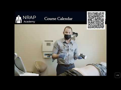
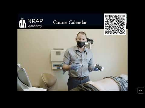
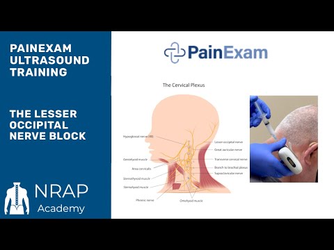
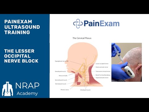
The video below, an excerpt from NRAP Academy’s Ultrasound Course, demonstrates the ultrasound-guided lesser occipital nerve block technique used in occipital neuralgia treatment, including transducer positioning, sonoanatomy of the posterior SCM border, and needle advancement.
If the video above doesn’t load, click the thumbnail to watch “The Ultrasound Guided Lesser Occipital Nerve Block” on YouTube.
Keep Learning
For a deeper discussion of occipital nerve anatomy and clinical pearls relevant to occipital neuralgia treatment, listen to “The Occipital Nerve” on The PainExam Podcast, where Dr. Rosenblum reviews the greater, lesser, and third occipital nerves. You can also find more ultrasound technique demonstrations on the NRAP Academy YouTube channel. For other ultrasound-guided nerve block techniques, see our posts on cervical radiculopathy and C6 nerve root injections and CRPS and the stellate ganglion block.
NRAP Academy Courses
Want to build hands-on skill with ultrasound-guided headache and peripheral nerve blocks? Explore the PainExam Pain Management Board Review and Refresher Course and the Ultrasound Training programs at NRAP Academy.
Looking for a Pain Specialist?
Looking for a pain specialist? Visit RosenblumPain.com or call (718) 436-7246. Patients outside the NYC/Brooklyn area can also book a virtual second-opinion consultation through RosenblumPain.com.
Disclaimer: This content is provided for general educational and informational purposes only and is intended for healthcare professionals, trainees, and patients seeking general information. It is not medical advice, does not constitute a diagnosis or treatment recommendation for any individual, and does not create a physician-patient relationship. Treatment decisions must always be individualized by a qualified, licensed healthcare provider based on a patient's specific history, examination, and current evidence-based guidelines. Dr. David Rosenblum, NRAP Academy, and PainExam make no warranties as to the completeness or applicability of this information and assume no liability for actions taken based on it. If you are a patient, please consult your own physician before making any medical decisions, and seek immediate medical attention for any urgent or emergency symptoms.